6QW3

Calcium-bound gelsolin domain 2


Experimental Data Snapshot

  • Method: X-RAY DIFFRACTION
  • Resolution: 1.30 Å
  • R-Value Free: 0.174 
  • R-Value Work: 0.154 
  • R-Value Observed: 0.156 

wwPDB Validation   3D Report Full Report


This is version 1.2 of the entry. See complete history


Literature

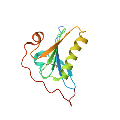
High-resolution crystal structure of gelsolin domain 2 in complex with the physiological calcium ion.

Bollati, M.Scalone, E.Boni, F.Mastrangelo, E.Giorgino, T.Milani, M.de Rosa, M.

(2019) Biochem Biophys Res Commun 518: 94-99

  • DOI: https://doi.org/10.1016/j.bbrc.2019.08.013
  • Primary Citation of Related Structures:  
    6QW3

  • PubMed Abstract: 

    The second domain of gelsolin (G2) hosts mutations responsible for a hereditary form of amyloidosis. The active form of gelsolin is Ca 2+ -bound; it is also a dynamic protein, hence structural biologists often rely on the study of the isolated G2. However, the wild type G2 structure that have been used so far in comparative studies is bound to a crystallographic Cd 2+ , in lieu of the physiological calcium. Here, we report the wild type structure of G2 in complex with Ca 2+ highlighting subtle ion-dependent differences. Previous findings on different G2 mutations are also briefly revised in light of these results.


  • Organizational Affiliation

    Istituto di Biofisica, Consiglio Nazionale delle Ricerche, Via Celoria 26, 20133, Milano, Italy; Dipartimento di Bioscienze, Università degli Studi di Milano, Via Celoria 26, 20133, Milano, Italy.


Macromolecules
Find similar proteins by:  (by identity cutoff)  |  3D Structure
Entity ID: 1
MoleculeChains Sequence LengthOrganismDetailsImage
Gelsolin119Homo sapiensMutation(s): 0 
Gene Names: GSN
UniProt & NIH Common Fund Data Resources
Find proteins for P06396 (Homo sapiens)
Explore P06396 
Go to UniProtKB:  P06396
PHAROS:  P06396
GTEx:  ENSG00000148180 
Entity Groups  
Sequence Clusters30% Identity50% Identity70% Identity90% Identity95% Identity100% Identity
UniProt GroupP06396
Sequence Annotations
Expand
  • Reference Sequence
Small Molecules
Ligands 1 Unique
IDChains Name / Formula / InChI Key2D Diagram3D Interactions
CA (Subject of Investigation/LOI)
Query on CA

Download Ideal Coordinates CCD File 
B [auth A]CALCIUM ION
Ca
BHPQYMZQTOCNFJ-UHFFFAOYSA-N
Experimental Data & Validation

Experimental Data

  • Method: X-RAY DIFFRACTION
  • Resolution: 1.30 Å
  • R-Value Free: 0.174 
  • R-Value Work: 0.154 
  • R-Value Observed: 0.156 
  • Space Group: P 2 21 21
Unit Cell:
Length ( Å )Angle ( ˚ )
a = 26.653α = 90
b = 50.275β = 90
c = 70.819γ = 90
Software Package:
Software NamePurpose
PHENIXrefinement
DIALSdata reduction
Aimlessdata scaling
PHASERphasing

Structure Validation

View Full Validation Report


Entry History & Funding Information

Deposition Data


Funding OrganizationLocationGrant Number
Amyloidosis FoundationUnited States--

Revision History  (Full details and data files)

  • Version 1.0: 2019-08-28
    Type: Initial release
  • Version 1.1: 2019-09-11
    Changes: Data collection, Database references
  • Version 1.2: 2024-01-24
    Changes: Data collection, Database references, Derived calculations, Refinement description