6PWT

Crystal structure of the cow C-type carbohydrate-recognition domain of CD23 in the presence of GlcNAc2Man3 oligosaccharide


Experimental Data Snapshot

  • Method: X-RAY DIFFRACTION
  • Resolution: 2.70 Å
  • R-Value Free: 0.251 
  • R-Value Work: 0.199 
  • R-Value Observed: 0.201 

wwPDB Validation   3D Report Full Report


This is version 2.1 of the entry. See complete history


Literature

CD23 is a glycan-binding receptor in some mammalian species.

Jegouzo, S.A.F.Feinberg, H.Morrison, A.G.Holder, A.May, A.Huang, Z.Jiang, L.Lasanajak, Y.Smith, D.F.Werling, D.Drickamer, K.Weis, W.I.Taylor, M.E.

(2019) J Biol Chem 294: 14845-14859

  • DOI: https://doi.org/10.1074/jbc.RA119.010572
  • Primary Citation of Related Structures:  
    6PWR, 6PWS, 6PWT

  • PubMed Abstract: 

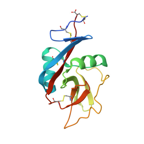
    CD23, the low-affinity IgE receptor found on B lymphocytes and other cells, contains a C-terminal lectin-like domain that resembles C-type carbohydrate-recognition domains (CRDs) found in many glycan-binding receptors. In most mammalian species, the CD23 residues required to form a sugar-binding site are present, although binding of CD23 to IgE does not involve sugars. Solid-phase binding competition assays, glycoprotein blotting experiments, and glycan array analysis employing the lectin-like domains of cow and mouse CD23 demonstrate that they bind to mannose, GlcNAc, glucose, and fucose and to glycoproteins that bear these sugars in nonreducing terminal positions. Crystal structures of the cow CRD in the presence of α-methyl mannoside and GlcNAcβ1-2Man reveal that a range of oligosaccharide ligands can be accommodated in an open binding site in which most interactions are with a single terminal sugar residue. Although mouse CD23 shows a pattern of monosaccharide and glycoprotein binding similar to cow CD23, the binding is weaker. In contrast, no sugar binding was observed in similar experiments with human CD23. The absence of sugar-binding activity correlates with accumulation of mutations in the gene for CD23 in the primate lineage leading to humans, resulting in loss of key sugar-binding residues. These results are consistent with a role for CD23 in many species as a receptor for potentially pathogenic microorganisms as well as IgE. However, the ability of CD23 to bind several different ligands varies between species, suggesting that it has distinct functions in different organisms.


  • Organizational Affiliation

    Department of Life Sciences, Imperial College London, London SW7 2AZ, United Kingdom.


Macromolecules
Find similar proteins by:  (by identity cutoff)  |  3D Structure
Entity ID: 1
MoleculeChains Sequence LengthOrganismDetailsImage
Fc fragment of IgE receptor II134Bos taurusMutation(s): 0 
Gene Names: FCER2
UniProt
Find proteins for E1BIQ4 (Bos taurus)
Explore E1BIQ4 
Go to UniProtKB:  E1BIQ4
Entity Groups  
Sequence Clusters30% Identity50% Identity70% Identity90% Identity95% Identity100% Identity
UniProt GroupE1BIQ4
Sequence Annotations
Expand
  • Reference Sequence
Oligosaccharides

Help

Entity ID: 2
MoleculeChains Length2D Diagram Glycosylation3D Interactions
2-acetamido-2-deoxy-beta-D-glucopyranose-(1-2)-alpha-D-mannopyranose-(1-3)-[2-acetamido-2-deoxy-beta-D-glucopyranose-(1-2)-alpha-D-mannopyranose-(1-6)]alpha-D-mannopyranose
B
5N/A
Glycosylation Resources
GlyTouCan:  G19266OP
GlyCosmos:  G19266OP
GlyGen:  G19266OP
Experimental Data & Validation

Experimental Data

  • Method: X-RAY DIFFRACTION
  • Resolution: 2.70 Å
  • R-Value Free: 0.251 
  • R-Value Work: 0.199 
  • R-Value Observed: 0.201 
  • Space Group: P 31 2 1
Unit Cell:
Length ( Å )Angle ( ˚ )
a = 33.167α = 90
b = 33.167β = 90
c = 207.814γ = 120
Software Package:
Software NamePurpose
XDSdata reduction
Aimlessdata scaling
PHASERphasing
PHENIXrefinement
PDB_EXTRACTdata extraction

Structure Validation

View Full Validation Report


Entry History & Funding Information

Deposition Data


Funding OrganizationLocationGrant Number
Biotechnology and Biological Sciences Research Council (BBSRC)United KingdomBB/P005659/1

Revision History  (Full details and data files)

  • Version 1.0: 2019-09-11
    Type: Initial release
  • Version 1.1: 2019-09-18
    Changes: Data collection, Database references
  • Version 1.2: 2019-10-23
    Changes: Data collection, Database references
  • Version 1.3: 2020-01-01
    Changes: Author supporting evidence
  • Version 2.0: 2020-07-29
    Type: Remediation
    Reason: Carbohydrate remediation
    Changes: Atomic model, Data collection, Derived calculations, Structure summary
  • Version 2.1: 2023-10-11
    Changes: Data collection, Database references, Refinement description, Structure summary