


Crystal structure of a glucose-6-phosphate isomerase from Chlamydia trachomatis D/UW-3/Cx
Edwards, T.E., Dranow, D.M., Horanyi, P.S., Lorimer, D.D., Seattle Structural Genomics Center for Infectious DiseaseTo be published.
Experimental Data Snapshot
Entity ID: 1 | |||||
|---|---|---|---|---|---|
| Molecule | Chains | Sequence Length | Organism | Details | Image |
| Glucose-6-phosphate isomerase | 533 | Chlamydia trachomatis D/UW-3/CX | Mutation(s): 0 Gene Names: pgi_1, pgi, pgi_2, pgi_4, ERS095036_01728, ERS133246_04369, ERS133248_01345 EC: 5.3.1.9 | 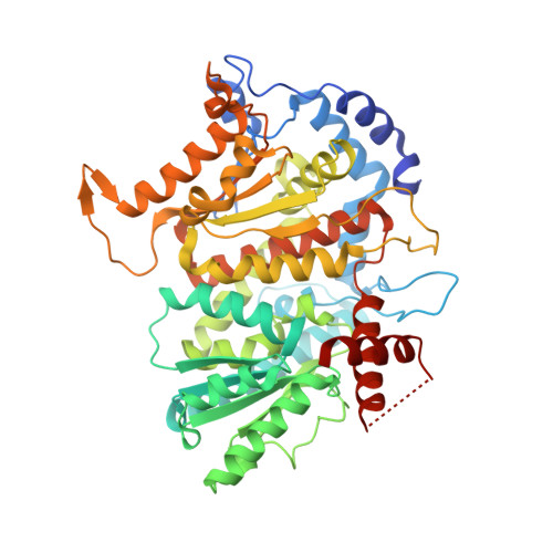 | |
Entity Groups | |||||
| Sequence Clusters | 30% Identity50% Identity70% Identity90% Identity95% Identity100% Identity | ||||
Sequence AnnotationsExpand | |||||
| |||||
| Ligands 2 Unique | |||||
|---|---|---|---|---|---|
| ID | Chains | Name / Formula / InChI Key | 2D Diagram | 3D Interactions | |
| G6Q (Subject of Investigation/LOI) Query on G6Q | E [auth A] | GLUCOSE-6-PHOSPHATE C6 H13 O9 P VFRROHXSMXFLSN-SLPGGIOYSA-N |  | ||
| ACT Query on ACT | B [auth A], C [auth A], D [auth A] | ACETATE ION C2 H3 O2 QTBSBXVTEAMEQO-UHFFFAOYSA-M |  | ||
| Length ( Å ) | Angle ( ˚ ) |
|---|---|
| a = 95.66 | α = 90 |
| b = 95.66 | β = 90 |
| c = 124.46 | γ = 90 |
| Software Name | Purpose |
|---|---|
| PHENIX | refinement |
| XDS | data reduction |
| XSCALE | data scaling |
| MOLREP | phasing |
| PDB_EXTRACT | data extraction |
RCSB PDB (citation) is hosted by
RCSB PDB is a member of the

