6OJ0

Cryo-EM reconstruction of Sulfolobus polyhedral virus 1 (SPV1)


Experimental Data Snapshot

  • Method: ELECTRON MICROSCOPY
  • Resolution: 3.70 Å
  • Aggregation State: PARTICLE 
  • Reconstruction Method: SINGLE PARTICLE 

wwPDB Validation   3D Report Full Report


This is version 1.5 of the entry. See complete history


Literature

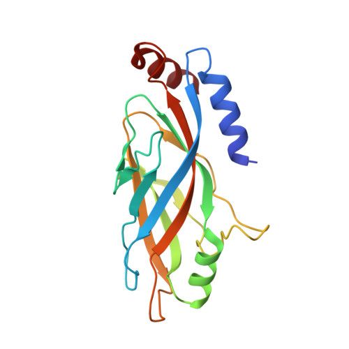
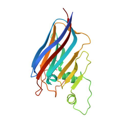
A packing for A-form DNA in an icosahedral virus.

Wang, F.Liu, Y.Su, Z.Osinski, T.de Oliveira, G.A.P.Conway, J.F.Schouten, S.Krupovic, M.Prangishvili, D.Egelman, E.H.

(2019) Proc Natl Acad Sci U S A 116: 22591-22597

  • DOI: https://doi.org/10.1073/pnas.1908242116
  • Primary Citation of Related Structures:  
    6OJ0

  • PubMed Abstract: 

    Studies on viruses infecting archaea living in the most extreme environments continue to show a remarkable diversity of structures, suggesting that the sampling continues to be very sparse. We have used electron cryo-microscopy to study at 3.7-Å resolution the structure of the Sulfolobus polyhedral virus 1 (SPV1), which was originally isolated from a hot, acidic spring in Beppu, Japan. The 2 capsid proteins with variant single jelly-roll folds form pentamers and hexamers which assemble into a T = 43 icosahedral shell. In contrast to tailed icosahedral double-stranded DNA (dsDNA) viruses infecting bacteria and archaea, and herpesviruses infecting animals and humans, where naked DNA is packed under very high pressure due to the repulsion between adjacent layers of DNA, the circular dsDNA in SPV1 is fully covered with a viral protein forming a nucleoprotein filament with attractive interactions between layers. Most strikingly, we have been able to show that the DNA is in an A-form, as it is in the filamentous viruses infecting hyperthermophilic acidophiles. Previous studies have suggested that DNA is in the B-form in bacteriophages, and our study is a direct visualization of the structure of DNA in an icosahedral virus.


  • Organizational Affiliation

    Department of Biochemistry and Molecular Genetics, University of Virginia, Charlottesville, VA 22908.


Macromolecules
Find similar proteins by:  (by identity cutoff)  |  3D Structure
Entity ID: 1
MoleculeChains Sequence LengthOrganismDetailsImage
Structural protein VP4181Sulfolobus polyhedral virus 1Mutation(s): 0 
UniProt
Find proteins for A0A1W6I187 (Sulfolobus polyhedral virus 1)
Explore A0A1W6I187 
Go to UniProtKB:  A0A1W6I187
Entity Groups  
Sequence Clusters30% Identity50% Identity70% Identity90% Identity95% Identity100% Identity
UniProt GroupA0A1W6I187
Sequence Annotations
Expand
  • Reference Sequence
Find similar proteins by:  (by identity cutoff)  |  3D Structure
Entity ID: 2
MoleculeChains Sequence LengthOrganismDetailsImage
Uncharacterized proteinQA [auth Z]187Sulfolobus polyhedral virus 1Mutation(s): 0 
UniProt
Find proteins for A0A1W6I162 (Sulfolobus polyhedral virus 1)
Explore A0A1W6I162 
Go to UniProtKB:  A0A1W6I162
Entity Groups  
Sequence Clusters30% Identity50% Identity70% Identity90% Identity95% Identity100% Identity
UniProt GroupA0A1W6I162
Sequence Annotations
Expand
  • Reference Sequence
Experimental Data & Validation

Experimental Data

  • Method: ELECTRON MICROSCOPY
  • Resolution: 3.70 Å
  • Aggregation State: PARTICLE 
  • Reconstruction Method: SINGLE PARTICLE 
EM Software:
TaskSoftware PackageVersion
RECONSTRUCTIONRELION

Structure Validation

View Full Validation Report


Entry History & Funding Information

Deposition Data


Funding OrganizationLocationGrant Number
National Institutes of Health/National Institute of General Medical Sciences (NIH/NIGMS)United StatesR35GM122510

Revision History  (Full details and data files)

  • Version 1.0: 2019-10-02
    Type: Initial release
  • Version 1.1: 2019-10-16
    Changes: Data collection, Derived calculations
  • Version 1.2: 2019-10-23
    Changes: Data collection, Database references
  • Version 1.3: 2019-11-06
    Changes: Data collection, Database references
  • Version 1.4: 2019-11-20
    Changes: Database references
  • Version 1.5: 2020-01-08
    Changes: Author supporting evidence