Influence of PEGylation on the Strength of Protein Surface Salt Bridges.
Xiao, Q., Draper, S.R.E., Smith, M.S., Brown, N., Pugmire, N.A.B., Ashton, D.S., Carter, A.J., Lawrence, E.E.K., Price, J.L.(2019) ACS Chem Biol 14: 1652-1659
- PubMed: 31188563
- DOI: https://doi.org/10.1021/acschembio.9b00432
- Primary Citation of Related Structures:
6O2E, 6O2F - PubMed Abstract:
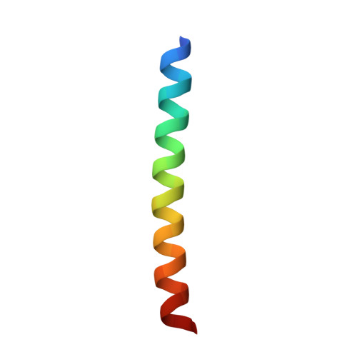
Conjugation of polyethylene glycol (PEGylation) is a well-known strategy for extending the serum half-life of protein drugs and for increasing their resistance to proteolysis and aggregation. We previously showed that PEGylation can increase protein conformational stability; the extent of PEG-based stabilization depends on the PEGylation site, the structure of the PEG-protein linker, and the ability of PEG to release water molecules from the surrounding protein surface to the bulk solvent. The strength of a noncovalent interaction within a protein depends strongly on its microenvironment, with salt-bridge and hydrogen-bond strength increasing in nonpolar versus aqueous environments. Accordingly, we wondered whether partial desolvation by PEG of the surrounding protein surface might result in measurable increases in the strength of a salt bridge near a PEGylation site. Here we explore this possibility using triple-mutant box analysis to assess the impact of PEGylation on the strength of nearby salt bridges at specific locations within three peptide model systems. The results indicate that PEG can increase the nearby salt-bridge strength, though this effect is not universal, and its precise structural prerequisites are not a simple function of secondary structural context, of the orientation and distance between the PEGylation site and salt bridge, or of salt-bridge residue identity. We obtained high-resolution X-ray diffraction data for a PEGylated peptide in which PEG enhances the strength of a nearby salt bridge. Comparing the electron density map of this PEGylated peptide versus that of its non-PEGylated counterpart provides evidence of localized protein surface desolvation as a mechanism for PEG-based salt-bridge stabilization.
Organizational Affiliation:
Department of Chemistry and Biochemistry , Brigham Young University , Provo , Utah 84602 , United States.