6NVY

Crystal structure of penicillin G acylase from Bacillus thermotolerans


Experimental Data Snapshot

  • Method: X-RAY DIFFRACTION
  • Resolution: 2.27 Å
  • R-Value Free: 0.200 
  • R-Value Work: 0.172 
  • R-Value Observed: 0.174 

wwPDB Validation   3D Report Full Report


This is version 1.2 of the entry. See complete history


Literature

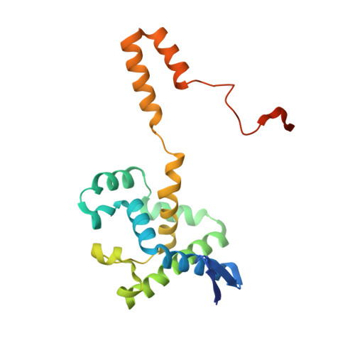
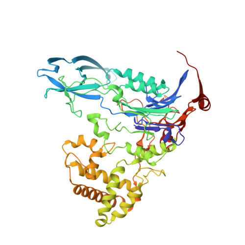
Crystal structures and protein engineering of three different penicillin G acylases from Gram-positive bacteria with different thermostability.

Mayer, J.Pippel, J.Gunther, G.Muller, C.Lauermann, A.Knuuti, T.Blankenfeldt, W.Jahn, D.Biedendieck, R.

(2019) Appl Microbiol Biotechnol 103: 7537-7552

  • DOI: https://doi.org/10.1007/s00253-019-09977-8
  • Primary Citation of Related Structures:  
    6NVW, 6NVX, 6NVY

  • PubMed Abstract: 

    Penicillin G acylase (PGA) catalyzes the hydrolysis of penicillin G to 6-aminopenicillanic acid and phenylacetic acid, which provides the precursor for most semisynthetic penicillins. Most applications rely on PGAs from Gram-negative bacteria. Here we describe the first three crystal structures for PGAs from Gram-positive Bacilli and their utilization in protein engineering experiments for the manipulation of their thermostability. PGAs from Bacillus megaterium (BmPGA, T m  = 56.0 °C), Bacillus thermotolerans (BtPGA, T m  = 64.5 °C), and Bacillus sp. FJAT-27231 (FJAT-PGA, T m  = 74.3 °C) were recombinantly produced with B. megaterium, secreted, purified to apparent heterogeneity, and crystallized. Structures with resolutions of 2.20 Å (BmPGA), 2.27 Å (BtPGA), and 1.36 Å (FJAT-PGA) were obtained. They revealed high overall similarity, reflecting the high identity of up to approx. 75%. Notably, the active center displays a deletion of more than ten residues with respect to PGAs from Gram-negatives. This enlarges the substrate binding site and may indicate a different substrate spectrum. Based on the structures, ten single-chain FJAT-PGAs carrying artificial linkers were produced. However, in all cases, complete linker cleavage was observed. While thermostability remained in the wild-type range, the enzymatic activity dropped between 30 and 60%. Furthermore, four hybrid PGAs carrying subunits from two different enzymes were successfully produced. Their thermostabilities mostly lay between the values of the two mother enzymes. For one PGA increased, enzyme activity was observed. Overall, the three novel PGA structures combined with initial protein engineering experiments provide the basis for establishment of new PGA-based biotechnological processes.


  • Organizational Affiliation

    Institute of Microbiology and Braunschweig Integrated Centre of Systems Biology (BRICS), Technische Universität Braunschweig, Rebenring 56, 38106, Braunschweig, Germany.


Macromolecules
Find similar proteins by:  (by identity cutoff)  |  3D Structure
Entity ID: 1
MoleculeChains Sequence LengthOrganismDetailsImage
Penicillin G acylase
A, C
212Bacillus thermotoleransMutation(s): 0 
Gene Names: QY95_01219
UniProt
Find proteins for A0A0F5I5V4 (Bacillus thermotolerans)
Explore A0A0F5I5V4 
Go to UniProtKB:  A0A0F5I5V4
Entity Groups  
Sequence Clusters30% Identity50% Identity70% Identity90% Identity95% Identity100% Identity
UniProt GroupA0A0F5I5V4
Sequence Annotations
Expand
  • Reference Sequence
Find similar proteins by:  (by identity cutoff)  |  3D Structure
Entity ID: 2
MoleculeChains Sequence LengthOrganismDetailsImage
Penicillin G acylase
B, D
538Bacillus thermotoleransMutation(s): 0 
Gene Names: QY95_01219
UniProt
Find proteins for A0A0F5I5V4 (Bacillus thermotolerans)
Explore A0A0F5I5V4 
Go to UniProtKB:  A0A0F5I5V4
Entity Groups  
Sequence Clusters30% Identity50% Identity70% Identity90% Identity95% Identity100% Identity
UniProt GroupA0A0F5I5V4
Sequence Annotations
Expand
  • Reference Sequence
Small Molecules
Ligands 2 Unique
IDChains Name / Formula / InChI Key2D Diagram3D Interactions
GOL
Query on GOL

Download Ideal Coordinates CCD File 
K [auth B]GLYCEROL
C3 H8 O3
PEDCQBHIVMGVHV-UHFFFAOYSA-N
CA
Query on CA

Download Ideal Coordinates CCD File 
E [auth A]
F [auth B]
G [auth B]
H [auth B]
I [auth B]
E [auth A],
F [auth B],
G [auth B],
H [auth B],
I [auth B],
J [auth B],
L [auth C],
M [auth D],
N [auth D],
O [auth D],
P [auth D],
Q [auth D]
CALCIUM ION
Ca
BHPQYMZQTOCNFJ-UHFFFAOYSA-N
Experimental Data & Validation

Experimental Data

  • Method: X-RAY DIFFRACTION
  • Resolution: 2.27 Å
  • R-Value Free: 0.200 
  • R-Value Work: 0.172 
  • R-Value Observed: 0.174 
  • Space Group: H 3
Unit Cell:
Length ( Å )Angle ( ˚ )
a = 172.996α = 90
b = 172.996β = 90
c = 209.566γ = 120
Software Package:
Software NamePurpose
XDSdata reduction
Aimlessdata scaling
PHASERphasing
PHENIXrefinement
PDB_EXTRACTdata extraction

Structure Validation

View Full Validation Report


Entry History & Funding Information

Deposition Data


Funding OrganizationLocationGrant Number
German Research FoundationGermanySPP1934

Revision History  (Full details and data files)

  • Version 1.0: 2019-07-03
    Type: Initial release
  • Version 1.1: 2019-09-11
    Changes: Data collection, Database references
  • Version 1.2: 2024-01-24
    Changes: Data collection, Database references, Derived calculations, Refinement description