6NFY

Crystal structure of nonphosphorylated, HPK1 kinase domain in complex with sunitinib in the inactive state.


Experimental Data Snapshot

  • Method: X-RAY DIFFRACTION
  • Resolution: 2.17 Å
  • R-Value Free: 0.219 
  • R-Value Work: 0.192 
  • R-Value Observed: 0.194 

wwPDB Validation   3D Report Full Report


Ligand Structure Quality Assessment 


This is version 1.4 of the entry. See complete history


Literature

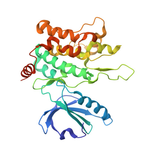
Multiple conformational states of the HPK1 kinase domain in complex with sunitinib reveal the structural changes accompanying HPK1 trans-regulation.

Johnson, E.McTigue, M.Gallego, R.A.Johnson, T.W.Timofeevski, S.Maestre, M.Fisher, T.S.Kania, R.Sawasdikosol, S.Burakoff, S.Cronin, C.N.

(2019) J Biol Chem 294: 9029-9036

  • DOI: https://doi.org/10.1074/jbc.AC119.007466
  • Primary Citation of Related Structures:  
    6NFY, 6NFZ, 6NG0

  • PubMed Abstract: 

    Hematopoietic progenitor kinase 1 (HPK1 or MAP4K1) is a Ser/Thr kinase that operates via the c-Jun N-terminal kinase (JNK) and extracellular signal-regulated kinase (ERK) signaling pathways to dampen the T-cell response and antitumor immunity. Accordingly, selective HPK1 inhibition is considered a means to enhance antitumor immunity. Sunitinib, a multi-receptor tyrosine kinase (RTK) inhibitor approved for the management of gastrointestinal stromal tumors (GISTs), renal cell carcinoma (RCC), and pancreatic cancer, has been reported to inhibit HPK1 in vitro In this report, we describe the crystal structures of the native HPK1 kinase domain in both nonphosphorylated and doubly phosphorylated states, in addition to a double phosphomimetic mutant (T165E,S171E), each complexed with sunitinib at 2.17-3.00-Å resolutions. The native nonphosphorylated cocrystal structure revealed an inactive dimer in which the activation loop of each monomer partially occupies the ATP- and substrate-binding sites of the partner monomer. In contrast, the structure of the protein with a doubly phosphorylated activation loop exhibited an active kinase conformation with a greatly reduced monomer-monomer interface. Conversely, the phosphomimetic mutant cocrystal structure disclosed an alternative arrangement in which the activation loops are in an extended domain-swapped configuration. These structural results indicate that HPK1 is a highly dynamic kinase that undergoes trans-regulation via dimer formation and extensive intramolecular and intermolecular remodeling of the activation segment.


  • Organizational Affiliation

    From the La Jolla Laboratories, Pfizer Worldwide Research and Development, San Diego, California 92121 and.


Macromolecules
Find similar proteins by:  (by identity cutoff)  |  3D Structure
Entity ID: 1
MoleculeChains Sequence LengthOrganismDetailsImage
Mitogen-activated protein kinase kinase kinase kinase 1
A, B
300Homo sapiensMutation(s): 0 
Gene Names: MAP4K1HPK1
EC: 2.7.11.1
UniProt & NIH Common Fund Data Resources
Find proteins for Q92918 (Homo sapiens)
Explore Q92918 
Go to UniProtKB:  Q92918
PHAROS:  Q92918
GTEx:  ENSG00000104814 
Entity Groups  
Sequence Clusters30% Identity50% Identity70% Identity90% Identity95% Identity100% Identity
UniProt GroupQ92918
Sequence Annotations
Expand
  • Reference Sequence
Small Molecules
Ligands 1 Unique
IDChains Name / Formula / InChI Key2D Diagram3D Interactions
B49
Query on B49

Download Ideal Coordinates CCD File 
C [auth A],
D [auth B]
N-[2-(diethylamino)ethyl]-5-[(Z)-(5-fluoro-2-oxo-1,2-dihydro-3H-indol-3-ylidene)methyl]-2,4-dimethyl-1H-pyrrole-3-carbo xamide
C22 H27 F N4 O2
WINHZLLDWRZWRT-ATVHPVEESA-N
Binding Affinity Annotations 
IDSourceBinding Affinity
B49 BindingDB:  6NFY Kd: 16 (nM) from 1 assay(s)
Binding MOAD:  6NFY Kd: 16 (nM) from 1 assay(s)
Experimental Data & Validation

Experimental Data

  • Method: X-RAY DIFFRACTION
  • Resolution: 2.17 Å
  • R-Value Free: 0.219 
  • R-Value Work: 0.192 
  • R-Value Observed: 0.194 
  • Space Group: H 3 2
Unit Cell:
Length ( Å )Angle ( ˚ )
a = 165.908α = 90
b = 165.908β = 90
c = 163.583γ = 120
Software Package:
Software NamePurpose
XDSdata reduction
Aimlessdata scaling
PHASERphasing
BUSTERrefinement
PDB_EXTRACTdata extraction

Structure Validation

View Full Validation Report


Ligand Structure Quality Assessment 


Entry History 

Deposition Data

Revision History  (Full details and data files)

  • Version 1.0: 2019-05-01
    Type: Initial release
  • Version 1.1: 2019-05-08
    Changes: Data collection, Database references
  • Version 1.2: 2019-06-19
    Changes: Data collection, Database references
  • Version 1.3: 2019-06-26
    Changes: Data collection, Database references
  • Version 1.4: 2024-03-13
    Changes: Data collection, Database references, Derived calculations, Structure summary