6MWS

crystal structure of the reduced Grx1 from Saccharomyces cerevisiae


Experimental Data Snapshot

  • Method: X-RAY DIFFRACTION
  • Resolution: 1.22 Å
  • R-Value Free: 0.172 
  • R-Value Work: 0.148 
  • R-Value Observed: 0.149 

wwPDB Validation   3D Report Full Report


This is version 1.1 of the entry. See complete history


Literature

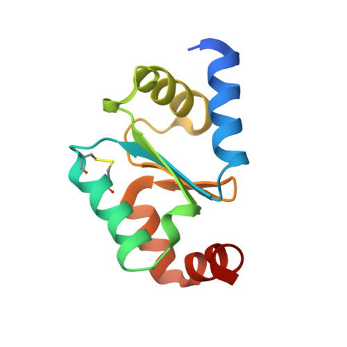
High-resolution crystal structure of the reduced Grx1 from Saccharomyces cerevisiae.

Maghool, S.La Fontaine, S.Maher, M.J.

(2019) Acta Crystallogr F Struct Biol Commun 75: 392-396

  • DOI: https://doi.org/10.1107/S2053230X19003327
  • Primary Citation of Related Structures:  
    6MWS

  • PubMed Abstract: 

    Grx1, a cytosolic thiol-disulfide oxidoreductase, actively maintains cellular redox homeostasis using glutathione substrates (reduced, GSH, and oxidized, GSSG). Here, the crystallization of reduced Grx1 from the yeast Saccharomyces cerevisiae (yGrx1) in space group P2 1 2 1 2 1 and its structure solution and refinement to 1.22 Å resolution are reported. To study the structure-function relationship of yeast Grx1, the crystal structure of reduced yGrx1 was compared with the existing structures of the oxidized and glutathionylated forms. These comparisons revealed structural differences in the conformations of residues neighbouring the Cys27-Cys30 active site which accompany alterations in the redox status of the protein.


  • Organizational Affiliation

    Department of Biochemistry and Genetics, La Trobe Institute for Molecular Science, La Trobe University, Melbourne, Victoria, Australia.


Macromolecules
Find similar proteins by:  (by identity cutoff)  |  3D Structure
Entity ID: 1
MoleculeChains Sequence LengthOrganismDetailsImage
Glutaredoxin-1115Saccharomyces cerevisiae S288CMutation(s): 0 
Gene Names: GRX1YCL035CYCL35C
UniProt
Find proteins for P25373 (Saccharomyces cerevisiae (strain ATCC 204508 / S288c))
Explore P25373 
Go to UniProtKB:  P25373
Entity Groups  
Sequence Clusters30% Identity50% Identity70% Identity90% Identity95% Identity100% Identity
UniProt GroupP25373
Sequence Annotations
Expand
  • Reference Sequence
Small Molecules
Ligands 1 Unique
IDChains Name / Formula / InChI Key2D Diagram3D Interactions
SO4
Query on SO4

Download Ideal Coordinates CCD File 
B [auth A]SULFATE ION
O4 S
QAOWNCQODCNURD-UHFFFAOYSA-L
Experimental Data & Validation

Experimental Data

  • Method: X-RAY DIFFRACTION
  • Resolution: 1.22 Å
  • R-Value Free: 0.172 
  • R-Value Work: 0.148 
  • R-Value Observed: 0.149 
  • Space Group: P 21 21 21
Unit Cell:
Length ( Å )Angle ( ˚ )
a = 41.364α = 90
b = 51.779β = 90
c = 57.193γ = 90
Software Package:
Software NamePurpose
REFMACrefinement
XDSdata reduction
Aimlessdata scaling
PHASERphasing
PDB_EXTRACTdata extraction

Structure Validation

View Full Validation Report


Entry History & Funding Information

Deposition Data


Funding OrganizationLocationGrant Number
Australian Research Council (ARC)AustraliaDP140102746

Revision History  (Full details and data files)

  • Version 1.0: 2019-05-15
    Type: Initial release
  • Version 1.1: 2020-01-01
    Changes: Author supporting evidence