De novo design of tunable, pH-driven conformational changes.
Boyken, S.E., Benhaim, M.A., Busch, F., Jia, M., Bick, M.J., Choi, H., Klima, J.C., Chen, Z., Walkey, C., Mileant, A., Sahasrabuddhe, A., Wei, K.Y., Hodge, E.A., Byron, S., Quijano-Rubio, A., Sankaran, B., King, N.P., Lippincott-Schwartz, J., Wysocki, V.H., Lee, K.K., Baker, D.(2019) Science 364: 658-664
- PubMed: 31097662
- DOI: https://doi.org/10.1126/science.aav7897
- Primary Citation of Related Structures:
6MSQ, 6MSR - PubMed Abstract:
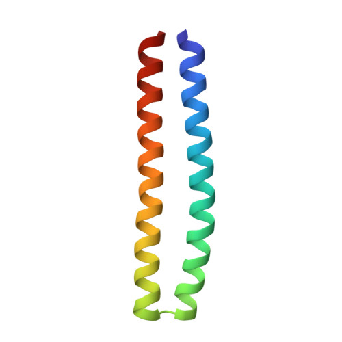
The ability of naturally occurring proteins to change conformation in response to environmental changes is critical to biological function. Although there have been advances in the de novo design of stable proteins with a single, deep free-energy minimum, the design of conformational switches remains challenging. We present a general strategy to design pH-responsive protein conformational changes by precisely preorganizing histidine residues in buried hydrogen-bond networks. We design homotrimers and heterodimers that are stable above pH 6.5 but undergo cooperative, large-scale conformational changes when the pH is lowered and electrostatic and steric repulsion builds up as the network histidine residues become protonated. The transition pH and cooperativity can be controlled through the number of histidine-containing networks and the strength of the surrounding hydrophobic interactions. Upon disassembly, the designed proteins disrupt lipid membranes both in vitro and after being endocytosed in mammalian cells. Our results demonstrate that environmentally triggered conformational changes can now be programmed by de novo protein design.
Organizational Affiliation:
Department of Biochemistry, University of Washington, Seattle, WA 98195, USA.