Structures of TOG1 and TOG2 from the human microtubule dynamics regulator CLASP1.
Leano, J.B., Slep, K.C.(2019) PLoS One 14: e0219823-e0219823
- PubMed: 31323070
- DOI: https://doi.org/10.1371/journal.pone.0219823
- Primary Citation of Related Structures:
6MQ5, 6MQ7 - PubMed Abstract:
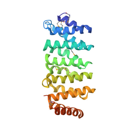
Tubulin-binding TOG domains are found arrayed in a number of proteins that regulate microtubule dynamics. While much is known about the structure and function of TOG domains from the XMAP215 microtubule polymerase family, less in known about the TOG domain array found in animal CLASP family members. The animal CLASP TOG array promotes microtubule pause, potentiates rescue, and limits catastrophe. How structurally distinct the TOG domains of animal CLASP are from one another, from XMAP215 family TOG domains, and whether a specific order of structurally distinct TOG domains in the TOG array is conserved across animal CLASP family members is poorly understood. We present the x-ray crystal structures of Homo sapiens (H.s.) CLASP1 TOG1 and TOG2. The structures of H.s. CLASP1 TOG1 and TOG2 are distinct from each other and from the previously determined structure of Mus musculus (M.m.) CLASP2 TOG3. Comparative analyses of CLASP family TOG domain structures determined to date across species and paralogs supports a conserved CLASP TOG array paradigm in which structurally distinct TOG domains are arrayed in a specific order. H.s. CLASP1 TOG1 bears structural similarity to the free-tubulin binding TOG domains of the XMAP215 family but lacks many of the key tubulin-binding determinants found in XMAP215 family TOG domains. This aligns with studies that report that animal CLASP family TOG1 domains cannot bind free tubulin or microtubules. In contrast, animal CLASP family TOG2 and TOG3 domains have reported microtubule-binding activity but are structurally distinct from the free-tubulin binding TOG domains of the XMAP215 family. H.s. CLASP1 TOG2 has a convex architecture, predicted to engage a hyper-curved tubulin state that may underlie its ability to limit microtubule catastrophe and promote rescue. M.m. CLASP2 TOG3 has unique structural elements in the C-terminal half of its α-solenoid domain that our modeling studies implicate in binding to laterally-associated tubulin subunits in the microtubule lattice in a mode similar to, yet distinct from those predicted for the XMAP215 family TOG4 domain. The potential ability of the animal CLASP family TOG3 domain to engage lateral tubulin subunits may underlie the microtubule rescue activity ascribed to the domain. These findings highlight the structural diversity of TOG domains within the CLASP family TOG array and provide a molecular foundation for understanding CLASP-dependent effects on microtubule dynamics.
Organizational Affiliation:
Department of Biochemistry & Biophysics, University of North Carolina, Chapel Hill, North Carolina, United States of America.