Functional Characterization and Structural Basis of an Efficient Di-C-glycosyltransferase fromGlycyrrhiza glabra.
Zhang, M., Li, F.D., Li, K., Wang, Z.L., Wang, Y.X., He, J.B., Su, H.F., Zhang, Z.Y., Chi, C.B., Shi, X.M., Yun, C.H., Zhang, Z.Y., Liu, Z.M., Zhang, L.R., Yang, D.H., Ma, M., Qiao, X., Ye, M.(2020) J Am Chem Soc 142: 3506-3512
- PubMed: 31986016
- DOI: https://doi.org/10.1021/jacs.9b12211
- Primary Citation of Related Structures:
6L5P, 6L5Q, 6L5R, 6L5S, 6L7H - PubMed Abstract:
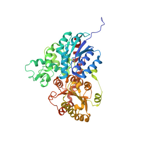
A highly efficient di- C -glycosyltransferase GgCGT was discovered from the medicinal plant Glycyrrhiza glabra . GgCGT catalyzes a two-step di- C -glycosylation of flopropione-containing substrates with conversion rates of >98%. To elucidate the catalytic mechanisms of GgCGT, we solved its crystal structures in complex with UDP-Glc, UDP-Gal, UDP/phloretin, and UDP/nothofagin, respectively. Structural analysis revealed that the sugar donor selectivity was controlled by the hydrogen-bond interactions of sugar hydroxyl groups with D390 and other key residues. The di- C -glycosylation capability of GgCGT was attributed to a spacious substrate-binding tunnel, and the G389K mutation could switch di- to mono- C -glycosylation. GgCGT is the first di- C -glycosyltransferase with a crystal structure, and the first C -glycosyltransferase with a complex structure containing a sugar acceptor. This work could benefit the development of efficient biocatalysts to synthesize C -glycosides with medicinal potential.
Organizational Affiliation:
State Key Laboratory of Natural and Biomimetic Drugs, School of Pharmaceutical Sciences , Peking University , 38 Xueyuan Road , Beijing 100191 , China.