6KBZ

Crystal structure of yedK with ssDNA containing a tetrahydrofuran abasic site


Experimental Data Snapshot

  • Method: X-RAY DIFFRACTION
  • Resolution: 1.65 Å
  • R-Value Free: 0.196 
  • R-Value Work: 0.160 
  • R-Value Observed: 0.162 

wwPDB Validation   3D Report Full Report


This is version 1.2 of the entry. See complete history


Literature

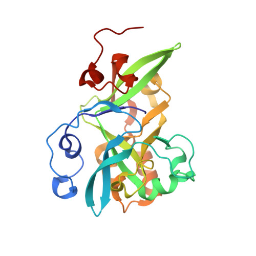
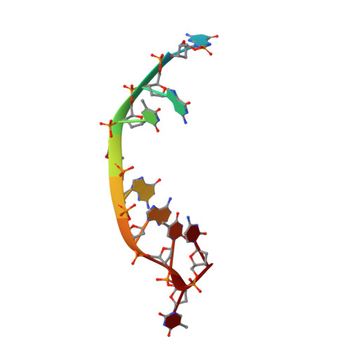
Molecular basis of abasic site sensing in single-stranded DNA by the SRAP domain of E. coli yedK.

Wang, N.Bao, H.Chen, L.Liu, Y.Li, Y.Wu, B.Huang, H.

(2019) Nucleic Acids Res 47: 10388-10399

  • DOI: https://doi.org/10.1093/nar/gkz744
  • Primary Citation of Related Structures:  
    6KBS, 6KBU, 6KBX, 6KBZ, 6KCQ, 6KIJ

  • PubMed Abstract: 

    HMCES and yedK were recently identified as sensors of abasic sites in ssDNA. In this study, we present multiple crystal structures captured in the apo-, nonspecific-substrate-binding, specific-substrate-binding, and product-binding states of yedK. In combination with biochemical data, we unveil the molecular basis of AP site sensing in ssDNA by yedK. Our results indicate that yedK has a strong preference for AP site-containing ssDNA over native ssDNA and that the conserved Glu105 residue is important for identifying AP sites in ssDNA. Moreover, our results reveal that a thiazolidine linkage is formed between yedK and AP sites in ssDNA, with the residues that stabilize the thiazolidine linkage important for the formation of DNA-protein crosslinks between yedK and the AP sites. We propose that our findings offer a unique platform to develop yedK and other SRAP domain-containing proteins as tools for detecting abasic sites in vitro and in vivo.


  • Organizational Affiliation

    Department of Biology, Southern University of Science and Technology, Shenzhen, Guangdong 518055, China.


Macromolecules

Find similar proteins by:  (by identity cutoff)  |  3D Structure
Entity ID: 1
MoleculeChains Sequence LengthOrganismDetailsImage
SOS response-associated proteinA [auth B],
C [auth D],
E [auth F],
G [auth H]
227Escherichia coliMutation(s): 0 
Gene Names: yedKC4J69_22885ECTO6_01993EFV06_12905EFV16_12155SAMEA3472108_01185SAMEA3752559_04370
EC: 3.4
UniProt
Find proteins for P76318 (Escherichia coli (strain K12))
Explore P76318 
Go to UniProtKB:  P76318
Entity Groups  
Sequence Clusters30% Identity50% Identity70% Identity90% Identity95% Identity100% Identity
UniProt GroupP76318
Sequence Annotations
Expand
  • Reference Sequence

Find similar nucleic acids by:  Sequence   |   3D Structure  

Entity ID: 2
MoleculeChains LengthOrganismImage
5'-D (*CP*GP*GP*TP* (3DR) P*GP*AP*TP*TP*C)-3'B [auth A],
D [auth C],
F [auth E],
H [auth G]
10Escherichia coli
Sequence Annotations
Expand
  • Reference Sequence
Experimental Data & Validation

Experimental Data

  • Method: X-RAY DIFFRACTION
  • Resolution: 1.65 Å
  • R-Value Free: 0.196 
  • R-Value Work: 0.160 
  • R-Value Observed: 0.162 
  • Space Group: P 1 21 1
Unit Cell:
Length ( Å )Angle ( ˚ )
a = 40.53α = 90
b = 111.391β = 96.14
c = 104.102γ = 90
Software Package:
Software NamePurpose
SCALEPACKdata scaling
PHENIXrefinement
PDB_EXTRACTdata extraction
HKL-3000data reduction
PHASERphasing

Structure Validation

View Full Validation Report


Entry History & Funding Information

Deposition Data


Funding OrganizationLocationGrant Number
Ministry of Science and Technology (China)China2018YFC1004500
National Natural Science Foundation of ChinaChinathe Thousand Young Talents Program
National Natural Science Foundation of ChinaChina31800619

Revision History  (Full details and data files)

  • Version 1.0: 2019-07-10
    Type: Initial release
  • Version 1.1: 2020-01-22
    Changes: Database references
  • Version 1.2: 2023-11-22
    Changes: Data collection, Database references, Derived calculations, Refinement description