6HYJ

PSPH Human phosphoserine phosphatase


Experimental Data Snapshot

  • Method: X-RAY DIFFRACTION
  • Resolution: 1.93 Å
  • R-Value Free: 0.263 
  • R-Value Work: 0.200 
  • R-Value Observed: 0.203 

wwPDB Validation   3D Report Full Report


Ligand Structure Quality Assessment 


This is version 2.2 of the entry. See complete history


Literature

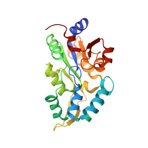
Crystal structures and snapshots along the reaction pathway of human phosphoserine phosphatase.

Haufroid, M.Mirgaux, M.Leherte, L.Wouters, J.

(2019) Acta Crystallogr D Struct Biol 75: 592-604

  • DOI: https://doi.org/10.1107/S2059798319006867
  • Primary Citation of Related Structures:  
    6HYJ, 6HYY, 6Q6J

  • PubMed Abstract: 

    The equilibrium between phosphorylation and dephosphorylation is one of the most important processes that takes place in living cells. Human phosphoserine phosphatase (hPSP) is a key enzyme in the production of serine by the dephosphorylation of phospho-L-serine. It is directly involved in the biosynthesis of other important metabolites such as glycine and D-serine (a neuromodulator). hPSP is involved in the survival mechanism of cancer cells and has recently been found to be an essential biomarker. Here, three new high-resolution crystal structures of hPSP (1.5-2.0 Å) in complexes with phosphoserine and with serine, which are the substrate and the product of the reaction, respectively, and in complex with a noncleavable substrate analogue (homocysteic acid) are presented. New types of interactions take place between the enzyme and its ligands. Moreover, the loop involved in the open/closed state of the enzyme is fully refined in a totally unfolded conformation. This loop is further studied through molecular-dynamics simulations. Finally, all of these analyses allow a more complete reaction mechanism for this enzyme to be proposed which is consistent with previous publications on the subject.


  • Organizational Affiliation

    Namur Medicine and Drug Innovation Center, Namur Research Institute for Life Science (NAMEDIC-NARILIS), Department of Chemistry, Laboratoire de Chimie Biologique Structurale (CBS), University of Namur (UNamur), 61 Rue de Bruxelles, 5000 Namur, Belgium.


Macromolecules
Find similar proteins by:  (by identity cutoff)  |  3D Structure
Entity ID: 1
MoleculeChains Sequence LengthOrganismDetailsImage
Phosphoserine phosphatase
A, B
224Homo sapiensMutation(s): 0 
Gene Names: PSPH
EC: 3.1.3.3
UniProt & NIH Common Fund Data Resources
Find proteins for P78330 (Homo sapiens)
Explore P78330 
Go to UniProtKB:  P78330
PHAROS:  P78330
GTEx:  ENSG00000146733 
Entity Groups  
Sequence Clusters30% Identity50% Identity70% Identity90% Identity95% Identity100% Identity
UniProt GroupP78330
Sequence Annotations
Expand
  • Reference Sequence
Experimental Data & Validation

Experimental Data

  • Method: X-RAY DIFFRACTION
  • Resolution: 1.93 Å
  • R-Value Free: 0.263 
  • R-Value Work: 0.200 
  • R-Value Observed: 0.203 
  • Space Group: C 2 2 21
Unit Cell:
Length ( Å )Angle ( ˚ )
a = 48.67α = 90
b = 128.97β = 90
c = 155.71γ = 90
Software Package:
Software NamePurpose
XSCALEdata scaling
PHENIXrefinement
PDB_EXTRACTdata extraction
XDSdata reduction
PHASERphasing

Structure Validation

View Full Validation Report


Ligand Structure Quality Assessment 


Entry History 

Deposition Data

Revision History  (Full details and data files)

  • Version 1.0: 2019-06-12
    Type: Initial release
  • Version 1.1: 2019-06-26
    Changes: Data collection, Database references
  • Version 2.0: 2019-10-02
    Changes: Advisory, Atomic model, Data collection, Database references, Derived calculations, Other, Polymer sequence, Source and taxonomy, Structure summary
  • Version 2.1: 2022-12-07
    Changes: Advisory, Database references, Derived calculations
  • Version 2.2: 2024-01-31
    Changes: Data collection, Refinement description