An Aromatic Dyad Motif in Dye Decolourising Peroxidases Has Implications for Free Radical Formation and Catalysis.
Chaplin, A.K., Chicano, T.M., Hampshire, B.V., Wilson, M.T., Hough, M.A., Svistunenko, D.A., Worrall, J.A.R.(2019) Chemistry 25: 6141-6153
- PubMed: 30945782
- DOI: https://doi.org/10.1002/chem.201806290
- Primary Citation of Related Structures:
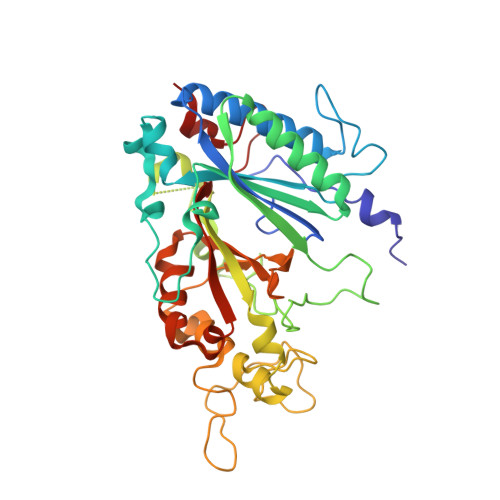
6GZW - PubMed Abstract:
Dye decolouring peroxidases (DyPs) are the most recent class of heme peroxidase to be discovered. On reacting with H 2 O 2 , DyPs form a high-valent iron(IV)-oxo species and a porphyrin radical (Compound I) followed by stepwise oxidation of an organic substrate. In the absence of substrate, the ferryl species decays to form transient protein-bound radicals on redox active amino acids. Identification of radical sites in DyPs has implications for their oxidative mechanism with substrate. Using a DyP from Streptomyces lividans, referred to as DtpA, which displays low reactivity towards synthetic dyes, activation with H 2 O 2 was explored. A Compound I EPR spectrum was detected, which in the absence of substrate decays to a protein-bound radical EPR signal. Using a newly developed version of the Tyrosyl Radical Spectra Simulation Algorithm, the radical EPR signal was shown to arise from a pristine tyrosyl radical and not a mixed Trp/Tyr radical that has been widely reported in DyP members exhibiting high activity with synthetic dyes. The radical site was identified as Tyr374, with kinetic studies inferring that although Tyr374 is not on the electron-transfer pathway from the dye RB19, its replacement with a Phe does severely compromise activity with other organic substrates. These findings hint at the possibility that alternative electron-transfer pathways for substrate oxidation are operative within the DyP family. In this context, a role for a highly conserved aromatic dyad motif is discussed.
Organizational Affiliation:
Present address: Department of Biochemistry, University of Cambridge, 80 Tennis Court Road, Cambridge, CB2 1GA, UK.