Structural evidence for an intransbase selection mechanism involving Loop1 in polymerase mu at an NHEJ double-strand break junction.
Loc'h, J., Gerodimos, C.A., Rosario, S., Tekpinar, M., Lieber, M.R., Delarue, M.(2019) J Biol Chem 294: 10579-10595
- PubMed: 31138645
- DOI: https://doi.org/10.1074/jbc.RA119.008739
- Primary Citation of Related Structures:
6GO3, 6GO4, 6GO5, 6GO6, 6GO7 - PubMed Abstract:
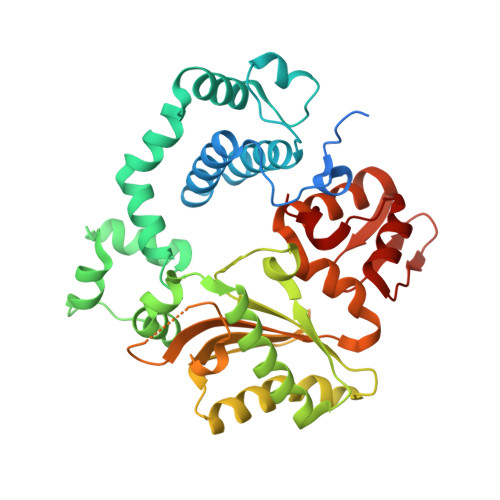
Eukaryotic DNA polymerase (Pol) X family members such as Pol μ and terminal deoxynucleotidyl transferase (TdT) are important components for the nonhomologous DNA end-joining (NHEJ) pathway. TdT participates in a specialized version of NHEJ, V(D)J recombination. It has primarily nontemplated polymerase activity but can take instructions across strands from the downstream dsDNA, and both activities are highly dependent on a structural element called Loop1. However, it is unclear whether Pol μ follows the same mechanism, because the structure of its Loop1 is disordered in available structures. Here, we used a chimeric TdT harboring Loop1 of Pol μ that recapitulated the functional properties of Pol μ in ligation experiments. We solved three crystal structures of this TdT chimera bound to several DNA substrates at 1.96-2.55 Å resolutions, including a full DNA double-strand break (DSB) synapsis. We then modeled the full Pol μ sequence in the context of one these complexes. The atomic structure of an NHEJ junction with a Pol X construct that mimics Pol μ in a reconstituted system explained the distinctive properties of Pol μ compared with TdT. The structure suggested a mechanism of base selection relying on Loop1 and taking instructions via the in trans templating base independently of the primer strand. We conclude that our atomic-level structural observations represent a paradigm shift for the mechanism of base selection in the Pol X family of DNA polymerases.
Organizational Affiliation:
From the Unité de Dynamique Structurale des Macromolécules, Institut Pasteur, UMR 3528 du CNRS, 25 Rue du Dr. Roux, 75015 Paris, France and.