6F92

Structure of the family GH92 alpha-mannosidase BT3965 from Bacteroides thetaiotaomicron in complex with Mannoimidazole (ManI)


Experimental Data Snapshot

  • Method: X-RAY DIFFRACTION
  • Resolution: 1.90 Å
  • R-Value Free: 0.208 
  • R-Value Work: 0.177 
  • R-Value Observed: 0.178 

wwPDB Validation   3D Report Full Report


Ligand Structure Quality Assessment 


This is version 1.2 of the entry. See complete history


Literature

Bacteroides thetaiotaomicron generates diverse alpha-mannosidase activities through subtle evolution of a distal substrate-binding motif.

Thompson, A.J.Spears, R.J.Zhu, Y.Suits, M.D.L.Williams, S.J.Gilbert, H.J.Davies, G.J.

(2018) Acta Crystallogr D Struct Biol 74: 394-404

  • DOI: https://doi.org/10.1107/S2059798318002942
  • Primary Citation of Related Structures:  
    6F8Z, 6F90, 6F91, 6F92

  • PubMed Abstract: 

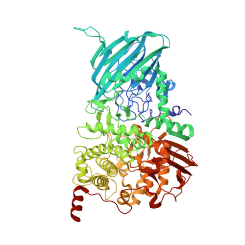
    A dominant human gut microbe, the well studied symbiont Bacteroides thetaiotaomicron (Bt), is a glyco-specialist that harbors a large repertoire of genes devoted to carbohydrate processing. Despite strong similarities among them, many of the encoded enzymes have evolved distinct substrate specificities, and through the clustering of cognate genes within operons termed polysaccharide-utilization loci (PULs) enable the fulfilment of complex biological roles. Structural analyses of two glycoside hydrolase family 92 α-mannosidases, BT3130 and BT3965, together with mechanistically relevant complexes at 1.8-2.5 Å resolution reveal conservation of the global enzyme fold and core catalytic apparatus despite different linkage specificities. Structure comparison shows that Bt differentiates the activity of these enzymes through evolution of a highly variable substrate-binding region immediately adjacent to the active site. These observations unveil a genetic/biochemical mechanism through which polysaccharide-processing bacteria can evolve new and specific biochemical activities from otherwise highly similar gene products.


  • Organizational Affiliation

    Department of Chemistry, University of York, Heslington, York YO10 5DD, England.


Macromolecules
Find similar proteins by:  (by identity cutoff)  |  3D Structure
Entity ID: 1
MoleculeChains Sequence LengthOrganismDetailsImage
Putative alpha-1,2-mannosidase
A, B, C, D
764Bacteroides thetaiotaomicronMutation(s): 0 
Gene Names: HMPREF2534_04919
UniProt
Find proteins for Q8A0Q6 (Bacteroides thetaiotaomicron (strain ATCC 29148 / DSM 2079 / JCM 5827 / CCUG 10774 / NCTC 10582 / VPI-5482 / E50))
Explore Q8A0Q6 
Go to UniProtKB:  Q8A0Q6
Entity Groups  
Sequence Clusters30% Identity50% Identity70% Identity90% Identity95% Identity100% Identity
UniProt GroupQ8A0Q6
Sequence Annotations
Expand
  • Reference Sequence
Small Molecules
Ligands 6 Unique
IDChains Name / Formula / InChI Key2D Diagram3D Interactions
MVL (Subject of Investigation/LOI)
Query on MVL

Download Ideal Coordinates CCD File 
H [auth A],
M [auth B],
S [auth C],
W [auth D]
(5R,6R,7S,8R)-5-(HYDROXYMETHYL)-5,6,7,8-TETRAHYDROIMIDAZO[1,2-A]PYRIDINE-6,7,8-TRIOL
C8 H12 N2 O4
RZRDQZQPTISYKY-JWXFUTCRSA-N
EDO
Query on EDO

Download Ideal Coordinates CCD File 
I [auth A],
N [auth B],
O [auth B],
X [auth D]
1,2-ETHANEDIOL
C2 H6 O2
LYCAIKOWRPUZTN-UHFFFAOYSA-N
NO3
Query on NO3

Download Ideal Coordinates CCD File 
Y [auth D],
Z [auth D]
NITRATE ION
N O3
NHNBFGGVMKEFGY-UHFFFAOYSA-N
CA
Query on CA

Download Ideal Coordinates CCD File 
F [auth A],
K [auth B],
Q [auth C],
U [auth D]
CALCIUM ION
Ca
BHPQYMZQTOCNFJ-UHFFFAOYSA-N
CL
Query on CL

Download Ideal Coordinates CCD File 
G [auth A],
L [auth B],
R [auth C],
V [auth D]
CHLORIDE ION
Cl
VEXZGXHMUGYJMC-UHFFFAOYSA-M
NA
Query on NA

Download Ideal Coordinates CCD File 
E [auth A],
J [auth B],
P [auth C],
T [auth D]
SODIUM ION
Na
FKNQFGJONOIPTF-UHFFFAOYSA-N
Binding Affinity Annotations 
IDSourceBinding Affinity
MVL BindingDB:  6F92 Ki: 6.00e+5 (nM) from 1 assay(s)
Experimental Data & Validation

Experimental Data

  • Method: X-RAY DIFFRACTION
  • Resolution: 1.90 Å
  • R-Value Free: 0.208 
  • R-Value Work: 0.177 
  • R-Value Observed: 0.178 
  • Space Group: P 1 21 1
Unit Cell:
Length ( Å )Angle ( ˚ )
a = 82.507α = 90
b = 186.904β = 91.74
c = 95.05γ = 90
Software Package:
Software NamePurpose
REFMACrefinement
XDSdata reduction
Aimlessdata scaling
PHASERphasing

Structure Validation

View Full Validation Report


Ligand Structure Quality Assessment 


Entry History & Funding Information

Deposition Data


Funding OrganizationLocationGrant Number
Biotechnology and Biological Sciences Research CouncilUnited KingdomBB/G016127/1
Royal SocietyUnited KingdomKen Murray Research Professorship

Revision History  (Full details and data files)

  • Version 1.0: 2018-05-02
    Type: Initial release
  • Version 1.1: 2018-05-09
    Changes: Data collection, Database references
  • Version 1.2: 2024-01-17
    Changes: Data collection, Database references, Derived calculations, Refinement description