6ERG

Complex of XLF and heterodimer Ku bound to DNA


Experimental Data Snapshot

  • Method: X-RAY DIFFRACTION
  • Resolution: 2.90 Å
  • R-Value Free: 0.244 
  • R-Value Work: 0.218 
  • R-Value Observed: 0.220 

wwPDB Validation   3D Report Full Report


This is version 1.2 of the entry. See complete history


Literature

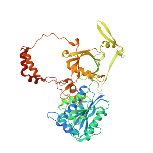
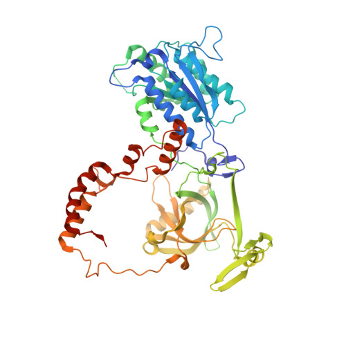
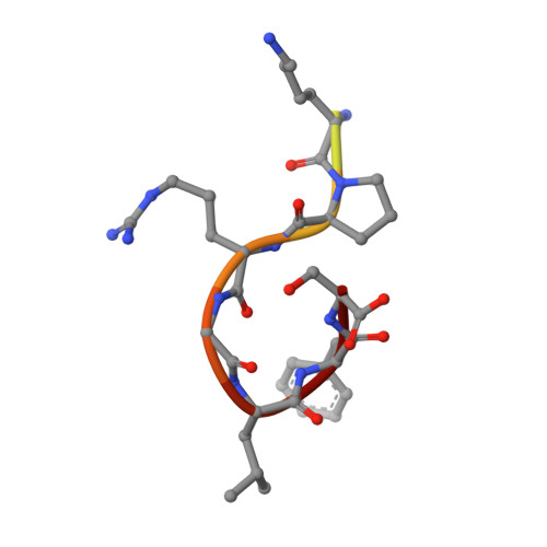
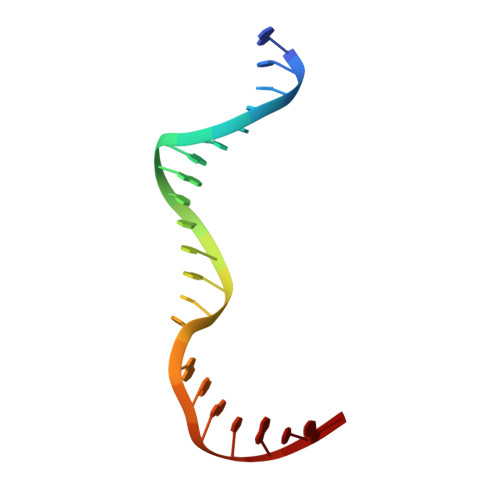
XLF and APLF bind Ku80 at two remote sites to ensure DNA repair by non-homologous end joining.

Nemoz, C.Ropars, V.Frit, P.Gontier, A.Drevet, P.Yu, J.Guerois, R.Pitois, A.Comte, A.Delteil, C.Barboule, N.Legrand, P.Baconnais, S.Yin, Y.Tadi, S.Barbet-Massin, E.Berger, I.Le Cam, E.Modesti, M.Rothenberg, E.Calsou, P.Charbonnier, J.B.

(2018) Nat Struct Mol Biol 25: 971-980

  • DOI: https://doi.org/10.1038/s41594-018-0133-6
  • Primary Citation of Related Structures:  
    6ERF, 6ERG, 6ERH

  • PubMed Abstract: 

    The Ku70-Ku80 (Ku) heterodimer binds rapidly and tightly to the ends of DNA double-strand breaks and recruits factors of the non-homologous end-joining (NHEJ) repair pathway through molecular interactions that remain unclear. We have determined crystal structures of the Ku-binding motifs (KBM) of the NHEJ proteins APLF (A-KBM) and XLF (X-KBM) bound to a Ku-DNA complex. The two KBM motifs bind remote sites of the Ku80 α/β domain. The X-KBM occupies an internal pocket formed by an unprecedented large outward rotation of the Ku80 α/β domain. We observe independent recruitment of the APLF-interacting protein XRCC4 and of XLF to laser-irradiated sites via binding of A- and X-KBMs, respectively, to Ku80. Finally, we show that mutation of the X-KBM and A-KBM binding sites in Ku80 compromises both the efficiency and accuracy of end joining and cellular radiosensitivity. A- and X-KBMs may represent two initial anchor points to build the intricate interaction network required for NHEJ.


  • Organizational Affiliation

    Institute for Integrative Biology of the Cell, Institute Joliot, CEA, CNRS, Université Paris-Sud, Université Paris-Saclay, Gif-sur-Yvette, France.


Macromolecules

Find similar proteins by:  (by identity cutoff)  |  3D Structure
Entity ID: 1
MoleculeChains Sequence LengthOrganismDetailsImage
X-ray repair cross-complementing protein 6
A, D
544Homo sapiensMutation(s): 0 
Gene Names: XRCC6G22P1
EC: 3.6.4 (PDB Primary Data), 4.2.99 (PDB Primary Data)
UniProt & NIH Common Fund Data Resources
Find proteins for P12956 (Homo sapiens)
Explore P12956 
Go to UniProtKB:  P12956
PHAROS:  P12956
GTEx:  ENSG00000196419 
Entity Groups  
Sequence Clusters30% Identity50% Identity70% Identity90% Identity95% Identity100% Identity
UniProt GroupP12956
Sequence Annotations
Expand
  • Reference Sequence
Find similar proteins by:  (by identity cutoff)  |  3D Structure
Entity ID: 2
MoleculeChains Sequence LengthOrganismDetailsImage
X-ray repair cross-complementing protein 5
B, E
572Homo sapiensMutation(s): 0 
Gene Names: XRCC5G22P2
EC: 3.6.4
UniProt & NIH Common Fund Data Resources
Find proteins for P13010 (Homo sapiens)
Explore P13010 
Go to UniProtKB:  P13010
PHAROS:  P13010
GTEx:  ENSG00000079246 
Entity Groups  
Sequence Clusters30% Identity50% Identity70% Identity90% Identity95% Identity100% Identity
UniProt GroupP13010
Sequence Annotations
Expand
  • Reference Sequence

Find similar proteins by:  Sequence   |   3D Structure  

Entity ID: 3
MoleculeChains Sequence LengthOrganismDetailsImage
Non-homologous end-joining factor 1
C, F
13Homo sapiensMutation(s): 0 
UniProt & NIH Common Fund Data Resources
Find proteins for Q9H9Q4 (Homo sapiens)
Explore Q9H9Q4 
Go to UniProtKB:  Q9H9Q4
PHAROS:  Q9H9Q4
GTEx:  ENSG00000187736 
Entity Groups  
Sequence Clusters30% Identity50% Identity70% Identity90% Identity95% Identity100% Identity
UniProt GroupQ9H9Q4
Sequence Annotations
Expand
  • Reference Sequence

Find similar nucleic acids by:  Sequence   |   3D Structure  

Entity ID: 4
MoleculeChains LengthOrganismImage
DNA (21-MER)G [auth H],
H [auth K]
21synthetic construct
Sequence Annotations
Expand
  • Reference Sequence
Find similar nucleic acids by:  (by identity cutoff)  |  3D Structure
Entity ID: 5
MoleculeChains LengthOrganismImage
DNA (34-MER)I [auth M],
J [auth R]
34synthetic construct
Sequence Annotations
Expand
  • Reference Sequence
Experimental Data & Validation

Experimental Data

  • Method: X-RAY DIFFRACTION
  • Resolution: 2.90 Å
  • R-Value Free: 0.244 
  • R-Value Work: 0.218 
  • R-Value Observed: 0.220 
  • Space Group: P 1 21 1
Unit Cell:
Length ( Å )Angle ( ˚ )
a = 111.71α = 90
b = 114.26β = 93.14
c = 127.17γ = 90
Software Package:
Software NamePurpose
XSCALEdata scaling
Cootmodel building
BUSTERrefinement
PDB_EXTRACTdata extraction
XDSdata reduction
MOLREPphasing

Structure Validation

View Full Validation Report


Entry History & Funding Information

Deposition Data


Funding OrganizationLocationGrant Number
INCAFrancePLBIO 2012-280
French National Research AgencyFranceANR-12-SVSE8-012

Revision History  (Full details and data files)

  • Version 1.0: 2018-10-17
    Type: Initial release
  • Version 1.1: 2019-04-03
    Changes: Data collection, Source and taxonomy
  • Version 1.2: 2024-01-17
    Changes: Advisory, Data collection, Database references, Refinement description