6EDD

Crystal structure of a GNAT Superfamily PA3944 acetyltransferase in complex with CoA (P1 space group)


Experimental Data Snapshot

  • Method: X-RAY DIFFRACTION
  • Resolution: 1.55 Å
  • R-Value Free: 0.194 
  • R-Value Work: 0.166 
  • R-Value Observed: 0.167 

wwPDB Validation   3D Report Full Report


Ligand Structure Quality Assessment 


This is version 1.4 of the entry. See complete history


Literature

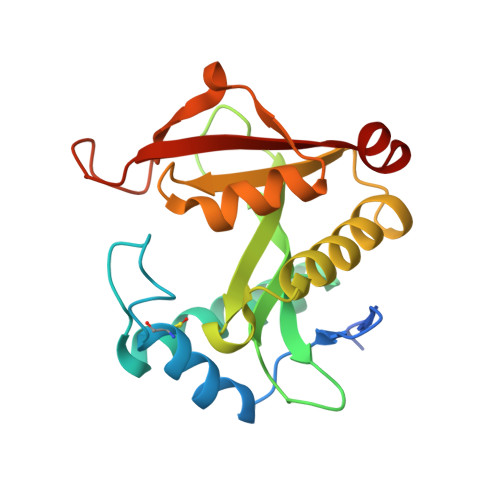
A Gcn5-Related N-Acetyltransferase (GNAT) Capable of Acetylating Polymyxin B and Colistin Antibiotics in Vitro.

Czub, M.P.Zhang, B.Chiarelli, M.P.Majorek, K.A.Joe, L.Porebski, P.J.Revilla, A.Wu, W.Becker, D.P.Minor, W.Kuhn, M.L.

(2018) Biochemistry 57: 7011-7020

  • DOI: https://doi.org/10.1021/acs.biochem.8b00946
  • Primary Citation of Related Structures:  
    6EDD, 6EDV

  • PubMed Abstract: 

    Deeper exploration of uncharacterized Gcn5-related N-acetyltransferases has the potential to expand our knowledge of the types of molecules that can be acylated by this important superfamily of enzymes and may offer new opportunities for biotechnological applications. While determining native or biologically relevant in vivo functions of uncharacterized proteins is ideal, their alternative or promiscuous in vitro capabilities provide insight into key active site interactions. Additionally, this knowledge can be exploited to selectively modify complex molecules and reduce byproducts when synthetic routes become challenging. During our exploration of uncharacterized Gcn5-related N-acetyltransferases from Pseudomonas aeruginosa, we identified such an example. We found that the PA3944 enzyme acetylates both polymyxin B and colistin on a single diaminobutyric acid residue closest to the macrocyclic ring of the antimicrobial peptide and determined the PA3944 crystal structure. This finding is important for several reasons. (1) To the best of our knowledge, this is the first report of enzymatic acylation of polymyxins and thus reveals a new type of substrate that this enzyme family can use. (2) The enzymatic acetylation offers a controlled method for antibiotic modification compared to classical promiscuous chemical methods. (3) The site of acetylation would reduce the overall positive charge of the molecule, which is important for reducing nephrotoxic effects and may be a salvage strategy for this important class of antibiotics. While the physiological substrate for this enzyme remains unknown, our structural and functional characterization of PA3944 offers insight into its unique noncanonical substrate specificity.


  • Organizational Affiliation

    Department of Molecular Physiology and Biological Physics , University of Virginia , Charlottesville , Virginia 22908 , United States.


Macromolecules
Find similar proteins by:  (by identity cutoff)  |  3D Structure
Entity ID: 1
MoleculeChains Sequence LengthOrganismDetailsImage
Acetyltransferase PA3944
A, B
194Pseudomonas aeruginosa PAO1Mutation(s): 0 
Gene Names: PA3944
EC: 2.3.1
UniProt
Find proteins for Q9HX72 (Pseudomonas aeruginosa (strain ATCC 15692 / DSM 22644 / CIP 104116 / JCM 14847 / LMG 12228 / 1C / PRS 101 / PAO1))
Explore Q9HX72 
Go to UniProtKB:  Q9HX72
Entity Groups  
Sequence Clusters30% Identity50% Identity70% Identity90% Identity95% Identity100% Identity
UniProt GroupQ9HX72
Sequence Annotations
Expand
  • Reference Sequence
Small Molecules
Ligands 4 Unique
IDChains Name / Formula / InChI Key2D Diagram3D Interactions
COA
Query on COA

Download Ideal Coordinates CCD File 
H [auth A],
O [auth B]
COENZYME A
C21 H36 N7 O16 P3 S
RGJOEKWQDUBAIZ-IBOSZNHHSA-N
TRS
Query on TRS

Download Ideal Coordinates CCD File 
I [auth B],
J [auth B]
2-AMINO-2-HYDROXYMETHYL-PROPANE-1,3-DIOL
C4 H12 N O3
LENZDBCJOHFCAS-UHFFFAOYSA-O
EDO
Query on EDO

Download Ideal Coordinates CCD File 
C [auth A],
D [auth A],
K [auth B]
1,2-ETHANEDIOL
C2 H6 O2
LYCAIKOWRPUZTN-UHFFFAOYSA-N
UNX
Query on UNX

Download Ideal Coordinates CCD File 
E [auth A]
F [auth A]
G [auth A]
L [auth B]
M [auth B]
E [auth A],
F [auth A],
G [auth A],
L [auth B],
M [auth B],
N [auth B]
UNKNOWN ATOM OR ION
X
Modified Residues  1 Unique
IDChains TypeFormula2D DiagramParent
CSX
Query on CSX
A, B
L-PEPTIDE LINKINGC3 H7 N O3 SCYS
Experimental Data & Validation

Experimental Data

  • Method: X-RAY DIFFRACTION
  • Resolution: 1.55 Å
  • R-Value Free: 0.194 
  • R-Value Work: 0.166 
  • R-Value Observed: 0.167 
  • Space Group: P 1
  • Diffraction Data: https://doi.org/10.18430/m36edd
Unit Cell:
Length ( Å )Angle ( ˚ )
a = 36.452α = 81.88
b = 44.175β = 73.32
c = 60.125γ = 89.94
Software Package:
Software NamePurpose
HKL-3000data reduction
HKL-3000data scaling
SCALEPACKdata scaling
HKL-3000phasing
MOLREPphasing
Cootmodel building
REFMACrefinement

Structure Validation

View Full Validation Report


Ligand Structure Quality Assessment 


Entry History & Funding Information

Deposition Data


Funding OrganizationLocationGrant Number
National Institutes of Health/National Institute Of Allergy and Infectious Diseases (NIH/NIAID)United States--

Revision History  (Full details and data files)

  • Version 1.0: 2018-08-22
    Type: Initial release
  • Version 1.1: 2018-12-19
    Changes: Data collection, Database references
  • Version 1.2: 2019-05-01
    Changes: Data collection, Database references
  • Version 1.3: 2019-12-18
    Changes: Author supporting evidence
  • Version 1.4: 2023-10-11
    Changes: Data collection, Database references, Refinement description