


Crystal Structure of a 1,2,4-Triazole Allosteric RNase H Inhibitor in Complex with HIV Reverse Transcriptase
Kirby, K.A., Sarafianos, S.G.To be published.
Experimental Data Snapshot
Entity ID: 1 | |||||
|---|---|---|---|---|---|
| Molecule | Chains | Sequence Length | Organism | Details | Image |
| Reverse transcriptase/ribonuclease H | 557 | Human immunodeficiency virus type 1 BH10 | Mutation(s): 1 EC: 3.1.13.2 | 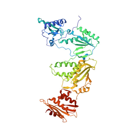 | |
UniProt | |||||
Find proteins for P03366 (Human immunodeficiency virus type 1 group M subtype B (isolate BH10)) Explore P03366 Go to UniProtKB: P03366 | |||||
Entity Groups | |||||
| Sequence Clusters | 30% Identity50% Identity70% Identity90% Identity95% Identity100% Identity | ||||
| UniProt Group | P03366 | ||||
Sequence AnnotationsExpand | |||||
| |||||
Entity ID: 2 | |||||
|---|---|---|---|---|---|
| Molecule | Chains | Sequence Length | Organism | Details | Image |
| Reverse transcriptase/ribonuclease H | 429 | Human immunodeficiency virus type 1 BH10 | Mutation(s): 1 EC: 3.1.13.2 | 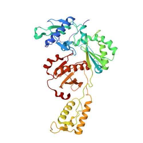 | |
UniProt | |||||
Find proteins for P03366 (Human immunodeficiency virus type 1 group M subtype B (isolate BH10)) Explore P03366 Go to UniProtKB: P03366 | |||||
Entity Groups | |||||
| Sequence Clusters | 30% Identity50% Identity70% Identity90% Identity95% Identity100% Identity | ||||
| UniProt Group | P03366 | ||||
Sequence AnnotationsExpand | |||||
| |||||
| Ligands 2 Unique | |||||
|---|---|---|---|---|---|
| ID | Chains | Name / Formula / InChI Key | 2D Diagram | 3D Interactions | |
| T90 Query on T90 | D [auth A] | 1-[4-methoxy-3-[[5-methyl-4-(phenylmethyl)-1,2,4-triazol-3-yl]sulfanylmethyl]phenyl]ethanone C20 H21 N3 O2 S GBQPKTFARABQAW-UHFFFAOYSA-N |  | ||
| MN Query on MN | C [auth A], E [auth A] | MANGANESE (II) ION Mn WAEMQWOKJMHJLA-UHFFFAOYSA-N |  | ||
| Length ( Å ) | Angle ( ˚ ) |
|---|---|
| a = 163.109 | α = 90 |
| b = 73.409 | β = 100.43 |
| c = 108.584 | γ = 90 |
| Software Name | Purpose |
|---|---|
| XDS | data reduction |
| Aimless | data scaling |
| PHASER | phasing |
| PHENIX | refinement |
| PDB_EXTRACT | data extraction |
| Funding Organization | Location | Grant Number |
|---|---|---|
| National Institutes of Health/National Institute Of Allergy and Infectious Diseases (NIH/NIAID) | United States | AI100890 |
RCSB PDB (citation) is hosted by
RCSB PDB is a member of the

