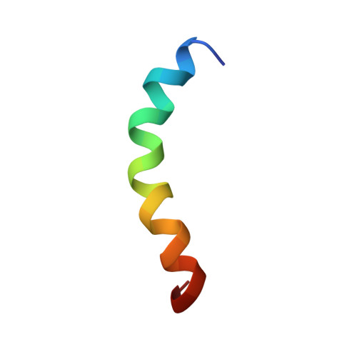
Helical Structure of Recombinant Melittin.
Ramirez, L.S., Pande, J., Shekhtman, A.(2019) J Phys Chem B 123: 356-368
- PubMed: 30570258
- DOI: https://doi.org/10.1021/acs.jpcb.8b08424
- Primary Citation of Related Structures:
6DST - PubMed Abstract:
Melittin is an extensively studied, 26-residue toxic peptide from honey bee venom. Because of its versatility in adopting a variety of secondary (helix or coil) and quaternary (monomer or tetramer) structures in various environments, melittin has been the focus of numerous investigations as a model peptide in protein folding studies as well as in studies involving binding to proteins, lipids, and polysaccharides. A significant body of evidence supports the view that melittin binds to these macromolecules in a predominantly helical conformation, but detailed structural knowledge of this conformation is lacking. In this report, we present nuclear magnetic resonance (NMR)-based structural insights into the helix formation of recombinant melittin in the presence of trifluoroethanol (TFE): a known secondary structure inducer in peptides. These studies were performed at neutral pH, with micromolar amounts of the peptide. Using nuclear Overhauser effect (NOE)-derived distance restraints from three-dimensional NMR spectra, we determined the atomic resolution solution NMR structure of recombinant melittin bearing a TFE-stabilized helix. To circumvent the complications with structure determination of small peptides with high conformational flexibility, we developed a workflow for enhancing proton NOEs by increasing the viscosity of the medium. In the TFE-containing medium, recombinant monomeric melittin forms a long, continuous helical structure, which consists of the N- and C-terminal α-helices and the noncanonical 3 10 -helix in the middle. The noncanonical 3 10 -helix is missing in the previously solved X-ray structure of tetrameric melittin and the NMR structure of melittin in methanol. Melittin's structure in TFE-containing medium provides insights into melittin's conformational transitions, which are relevant to the peptide's interactions with its biological targets.
Organizational Affiliation:
Department of Chemistry , State University of New York at Albany , Albany , New York 12222 , United States.