6D6I

Ube2V1 in complex with ubiquitin variant Ubv.V1.1 and Ube2N/Ubc13


Experimental Data Snapshot

  • Method: X-RAY DIFFRACTION
  • Resolution: 2.55 Å
  • R-Value Free: 0.296 
  • R-Value Work: 0.264 
  • R-Value Observed: 0.265 

wwPDB Validation   3D Report Full Report


This is version 1.4 of the entry. See complete history


Literature

Structural and Functional Analysis of Ubiquitin-based Inhibitors That Target the Backsides of E2 Enzymes.

Garg, P.Ceccarelli, D.F.Keszei, A.F.A.Kurinov, I.Sicheri, F.Sidhu, S.S.

(2020) J Mol Biol 432: 952-966

  • DOI: https://doi.org/10.1016/j.jmb.2019.09.024
  • Primary Citation of Related Structures:  
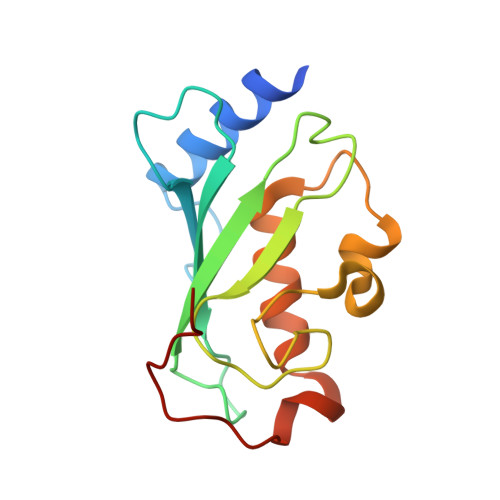
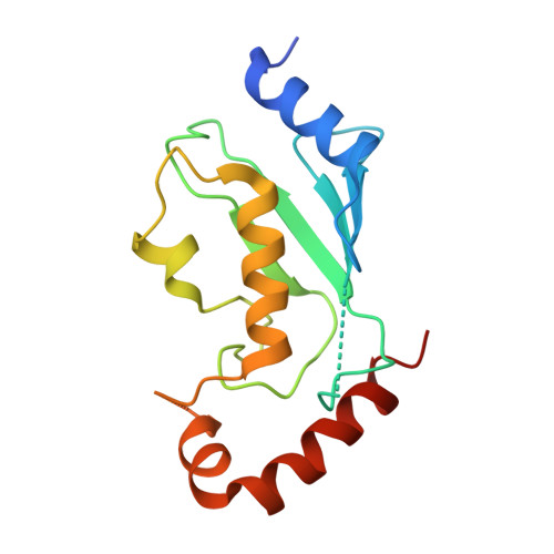
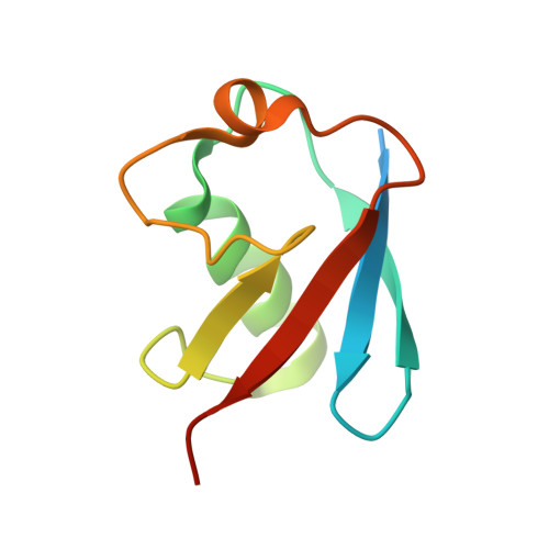
    6D4P, 6D68, 6D6I

  • PubMed Abstract: 

    Ubiquitin-conjugating E2 enzymes are central to the ubiquitination cascade and have been implicated in cancer and other diseases. Despite strong interest in developing specific E2 inhibitors, the shallow and exposed active site has proven recalcitrant to targeting with reversible small-molecule inhibitors. Here, we used phage display to generate highly potent and selective ubiquitin variants (UbVs) that target the E2 backside, which is located opposite to the active site. A UbV targeting Ube2D1 did not affect charging but greatly attenuated chain elongation. Likewise, a UbV targeting the E2 variant Ube2V1 did not interfere with the charging of its partner E2 enzyme but inhibited formation of diubiquitin. In contrast, a UbV that bound to the backside of Ube2G1 impeded the generation of thioester-linked ubiquitin to the active site cysteine of Ube2G1 by the E1 enzyme. Crystal structures of UbVs in complex with three E2 proteins revealed distinctive molecular interactions in each case, but they also highlighted a common backside pocket that the UbVs used for enhanced affinity and specificity. These findings validate the E2 backside as a target for inhibition and provide structural insights to aid inhibitor design and screening efforts.


  • Organizational Affiliation

    Department of Molecular Genetics, University of Toronto, Toronto, Ontario M5S 1A8, Canada; The Donnelly Centre for Cellular and Biomolecular Research, University of Toronto, 160 College Street, Toronto, Ontario M5S 3E1, Canada.


Macromolecules
Find similar proteins by:  (by identity cutoff)  |  3D Structure
Entity ID: 1
MoleculeChains Sequence LengthOrganismDetailsImage
Ubiquitin-conjugating enzyme E2 variant 1
A, D
143Homo sapiensMutation(s): 0 
Gene Names: UBE2V1CROC1UBE2VUEV1P/OKcl.19
UniProt & NIH Common Fund Data Resources
Find proteins for Q13404 (Homo sapiens)
Explore Q13404 
Go to UniProtKB:  Q13404
GTEx:  ENSG00000244687 
Entity Groups  
Sequence Clusters30% Identity50% Identity70% Identity90% Identity95% Identity100% Identity
UniProt GroupQ13404
Sequence Annotations
Expand
  • Reference Sequence
Find similar proteins by:  (by identity cutoff)  |  3D Structure
Entity ID: 2
MoleculeChains Sequence LengthOrganismDetailsImage
Ubiquitin-conjugating enzyme E2 N
B, E
155Homo sapiensMutation(s): 0 
Gene Names: UBE2NBLU
EC: 2.3.2.23
UniProt & NIH Common Fund Data Resources
Find proteins for P61088 (Homo sapiens)
Explore P61088 
Go to UniProtKB:  P61088
PHAROS:  P61088
GTEx:  ENSG00000177889 
Entity Groups  
Sequence Clusters30% Identity50% Identity70% Identity90% Identity95% Identity100% Identity
UniProt GroupP61088
Sequence Annotations
Expand
  • Reference Sequence
Find similar proteins by:  (by identity cutoff)  |  3D Structure
Entity ID: 3
MoleculeChains Sequence LengthOrganismDetailsImage
Ubv.V1.1
C, F
88Homo sapiensMutation(s): 8 
Gene Names: Uba52
UniProt
Find proteins for Q96H31 (Homo sapiens)
Explore Q96H31 
Go to UniProtKB:  Q96H31
Entity Groups  
Sequence Clusters30% Identity50% Identity70% Identity90% Identity95% Identity100% Identity
UniProt GroupQ96H31
Sequence Annotations
Expand
  • Reference Sequence
Experimental Data & Validation

Experimental Data

  • Method: X-RAY DIFFRACTION
  • Resolution: 2.55 Å
  • R-Value Free: 0.296 
  • R-Value Work: 0.264 
  • R-Value Observed: 0.265 
  • Space Group: P 32
Unit Cell:
Length ( Å )Angle ( ˚ )
a = 91.444α = 90
b = 91.444β = 90
c = 92.446γ = 120
Software Package:
Software NamePurpose
PHENIXrefinement
HKL-2000data reduction
HKL-2000data scaling
PHASERphasing
PDB_EXTRACTdata extraction

Structure Validation

View Full Validation Report


Entry History & Funding Information

Deposition Data


Funding OrganizationLocationGrant Number
Canadian Institutes of Health Research (CIHR)CanadaMOP-136956
Canadian Institutes of Health Research (CIHR)CanadaMOP-126129

Revision History  (Full details and data files)

  • Version 1.0: 2019-07-17
    Type: Initial release
  • Version 1.1: 2019-11-06
    Changes: Data collection, Database references
  • Version 1.2: 2020-01-08
    Changes: Author supporting evidence
  • Version 1.3: 2020-03-11
    Changes: Database references
  • Version 1.4: 2023-10-04
    Changes: Data collection, Database references, Refinement description