


6CQJ
Crystal structure of DR1 presenting the RQ13 peptide
- PDB DOI: https://doi.org/10.2210/pdb6CQJ/pdb
- Classification: IMMUNE SYSTEM
- Organism(s): Homo sapiens, HIV-1 M:B_HXB2R
- Expression System: Homo sapiens
- Mutation(s): No
- Deposited: 2018-03-15 Released: 2018-06-06
Experimental Data Snapshot
- Method: X-RAY DIFFRACTION
- Resolution: 2.75 Å
- R-Value Free: 0.249
- R-Value Work: 0.197
- R-Value Observed: 0.200

This is version 2.1 of the entry. See complete history.

Macromolecules
Find similar proteins by:
(by identity cutoff) | 3D Structure
Entity ID: 1 | |||||
|---|---|---|---|---|---|
| Molecule | Chains | Sequence Length | Organism | Details | Image |
| HLA class II histocompatibility antigen, DR alpha chain | 182 | Homo sapiens | Mutation(s): 0 Gene Names: HLA-DRA, HLA-DRA1 |  | |
UniProt & NIH Common Fund Data Resources | |||||
Find proteins for P01903 (Homo sapiens) Explore P01903 Go to UniProtKB: P01903 | |||||
PHAROS: P01903 GTEx: ENSG00000204287 | |||||
Entity Groups | |||||
| Sequence Clusters | 30% Identity50% Identity70% Identity90% Identity95% Identity100% Identity | ||||
| UniProt Group | P01903 | ||||
Sequence AnnotationsExpand | |||||
| |||||
Find similar proteins by:
(by identity cutoff) | 3D Structure
Entity ID: 2 | |||||
|---|---|---|---|---|---|
| Molecule | Chains | Sequence Length | Organism | Details | Image |
| HLA class II histocompatibility antigen, DRB1-1 beta chain | 189 | Homo sapiens | Mutation(s): 0 Gene Names: HLA-DRB1 | 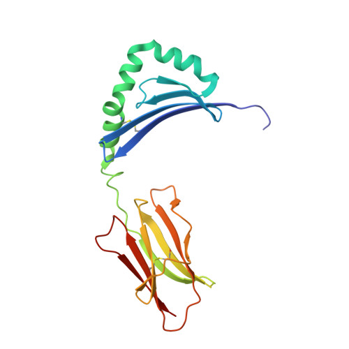 | |
UniProt & NIH Common Fund Data Resources | |||||
Find proteins for P01911 (Homo sapiens) Explore P01911 Go to UniProtKB: P01911 | |||||
PHAROS: P01911 GTEx: ENSG00000196126 | |||||
Entity Groups | |||||
| Sequence Clusters | 30% Identity50% Identity70% Identity90% Identity95% Identity100% Identity | ||||
| UniProt Group | P01911 | ||||
Sequence AnnotationsExpand | |||||
| |||||
Find similar proteins by: Sequence | 3D Structure
Entity ID: 3 | |||||
|---|---|---|---|---|---|
| Molecule | Chains | Sequence Length | Organism | Details | Image |
| Peptide from Capsid protein p24 | 13 | HIV-1 M:B_HXB2R | Mutation(s): 0 |  | |
UniProt | |||||
Find proteins for P04585 (Human immunodeficiency virus type 1 group M subtype B (isolate HXB2)) Explore P04585 Go to UniProtKB: P04585 | |||||
Entity Groups | |||||
| Sequence Clusters | 30% Identity50% Identity70% Identity90% Identity95% Identity100% Identity | ||||
| UniProt Group | P04585 | ||||
Sequence AnnotationsExpand | |||||
| |||||
Oligosaccharides
Entity ID: 4 | |||||
|---|---|---|---|---|---|
| Molecule | Chains | Length | 2D Diagram | Glycosylation | 3D Interactions |
| 2-acetamido-2-deoxy-beta-D-glucopyranose-(1-4)-2-acetamido-2-deoxy-beta-D-glucopyranose-(1-4)-2-acetamido-2-deoxy-beta-D-glucopyranose | J | 3 |  | N-Glycosylation | |
Glycosylation Resources | |||||
GlyTouCan: G47362BJ GlyCosmos: G47362BJ GlyGen: G47362BJ | |||||
Small Molecules
| Ligands 3 Unique | |||||
|---|---|---|---|---|---|
| ID | Chains | Name / Formula / InChI Key | 2D Diagram | 3D Interactions | |
| NAG Query on NAG | L [auth A], O [auth D] | 2-acetamido-2-deoxy-beta-D-glucopyranose C8 H15 N O6 OVRNDRQMDRJTHS-FMDGEEDCSA-N |  | ||
| PEG Query on PEG | K [auth A], M [auth B], P [auth E], Q [auth G] | DI(HYDROXYETHYL)ETHER C4 H10 O3 MTHSVFCYNBDYFN-UHFFFAOYSA-N |  | ||
| NA Query on NA | N [auth D], R [auth G] | SODIUM ION Na FKNQFGJONOIPTF-UHFFFAOYSA-N |  | ||
Biologically Interesting Molecules (External Reference) 1 Unique
Entity ID: 4 | |||||
|---|---|---|---|---|---|
| ID | Chains | Name | Type/Class | 2D Diagram | 3D Interactions |
| PRD_900017 Query on PRD_900017 | J | triacetyl-beta-chitotriose | Oligosaccharide / Inhibitor |  | |
Experimental Data & Validation
Experimental Data
- Method: X-RAY DIFFRACTION
- Resolution: 2.75 Å
- R-Value Free: 0.249
- R-Value Work: 0.197
- R-Value Observed: 0.200
- Space Group: P 1
Unit Cell:
| Length ( Å ) | Angle ( ˚ ) |
|---|---|
| a = 68.684 | α = 61.56 |
| b = 82.017 | β = 88.2 |
| c = 83.05 | γ = 86.47 |
| Software Name | Purpose |
|---|---|
| BUSTER | refinement |
| XDS | data reduction |
| Aimless | data scaling |
Entry History
Deposition Data
- Released Date: 2018-06-06 Deposition Author(s): Farenc, C., Gras, S., Rossjohn, J.
Revision History (Full details and data files)
- Version 1.0: 2018-06-06
Type: Initial release - Version 1.1: 2018-06-27
Changes: Data collection, Database references, Structure summary - Version 2.0: 2020-07-29
Type: Remediation
Reason: Carbohydrate remediation
Changes: Atomic model, Data collection, Derived calculations, Structure summary - Version 2.1: 2023-10-04
Changes: Data collection, Database references, Refinement description, Structure summary
