6A85

Crystal structure of a novel DNA quadruplex

  • Classification: DNA
  • Organism(s): synthetic construct
  • Mutation(s): No 

  • Deposited: 2018-07-06 Released: 2019-03-06 
  • Deposition Author(s): Liu, H.H., Gan, J.H.
  • Funding Organization(s): National Natural Science Foundation of China

Experimental Data Snapshot

  • Method: X-RAY DIFFRACTION
  • Resolution: 1.45 Å
  • R-Value Free: 0.169 
  • R-Value Work: 0.136 
  • R-Value Observed: 0.138 

wwPDB Validation   3D Report Full Report


This is version 1.1 of the entry. See complete history


Literature

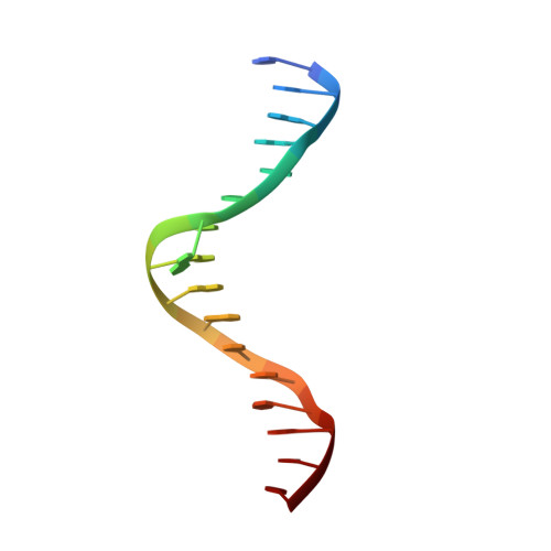
High-resolution DNA quadruplex structure containing all the A-, G-, C-, T-tetrads.

Liu, H.H.Wang, R.Yu, X.Shen, F.S.Lan, W.X.Haruehanroengra, P.Yao, Q.Q.Zhang, J.Chen, Y.Q.Li, S.H.Wu, B.X.Zheng, L.N.Ma, J.B.Lin, J.Z.Cao, C.Y.Li, J.X.Sheng, J.Gan, J.H.

(2018) Nucleic Acids Res 46: 11627-11638

  • DOI: https://doi.org/10.1093/nar/gky902
  • Primary Citation of Related Structures:  
    6A85

  • PubMed Abstract: 

    DNA can form diverse structures, which predefine their physiological functions. Besides duplexes that carry the genetic information, quadruplexes are the most well-studied DNA structures. In addition to their important roles in recombination, replication, transcription and translation, DNA quadruplexes have also been applied as diagnostic aptamers and antidisease therapeutics. Herein we further expand the sequence and structure complexity of DNA quadruplex by presenting a high-resolution crystal structure of DNA1 (5'-AGAGAGATGGGTGCGTT-3'). This is the first quadruplex structure that contains all the internal A-, G-, C-, T-tetrads, A:T:A:T tetrads and bulged nucleotides in one single structure; as revealed by site-specific mutagenesis and biophysical studies, the central ATGGG motif plays important role in the quadruplex formation. Interestingly, our structure also provides great new insights into cation recognition, including the first-time reported Pb2+, by tetrad structures.


  • Organizational Affiliation

    State Key Laboratory of Genetic Engineering, Collaborative Innovation Center of Genetics and Development, Department of Physiology and Biophysics, School of Life Sciences, Fudan University, Shanghai 200433, China.


Macromolecules

Find similar nucleic acids by:  Sequence   |   3D Structure  

Entity ID: 1
MoleculeChains LengthOrganismImage
DNA (5'-D(*AP*GP*AP*GP*AP*GP*AP*TP*GP*GP*GP*TP*GP*CP*GP*TP*T)-3')
A, B
17synthetic construct
Sequence Annotations
Expand
  • Reference Sequence
Small Molecules
Ligands 4 Unique
IDChains Name / Formula / InChI Key2D Diagram3D Interactions
PB
Query on PB

Download Ideal Coordinates CCD File 
N [auth A]LEAD (II) ION
Pb
RVPVRDXYQKGNMQ-UHFFFAOYSA-N
NA
Query on NA

Download Ideal Coordinates CCD File 
E [auth A]
F [auth A]
G [auth A]
H [auth A]
I [auth A]
E [auth A],
F [auth A],
G [auth A],
H [auth A],
I [auth A],
J [auth A],
K [auth A],
L [auth A],
M [auth A],
O [auth A],
P [auth B]
SODIUM ION
Na
FKNQFGJONOIPTF-UHFFFAOYSA-N
NH4
Query on NH4

Download Ideal Coordinates CCD File 
Q [auth B]AMMONIUM ION
H4 N
QGZKDVFQNNGYKY-UHFFFAOYSA-O
LI
Query on LI

Download Ideal Coordinates CCD File 
C [auth A],
D [auth A]
LITHIUM ION
Li
HBBGRARXTFLTSG-UHFFFAOYSA-N
Experimental Data & Validation

Experimental Data

  • Method: X-RAY DIFFRACTION
  • Resolution: 1.45 Å
  • R-Value Free: 0.169 
  • R-Value Work: 0.136 
  • R-Value Observed: 0.138 
  • Space Group: P 21 21 2
Unit Cell:
Length ( Å )Angle ( ˚ )
a = 37.822α = 90
b = 46.651β = 90
c = 51.311γ = 90
Software Package:
Software NamePurpose
PHENIXrefinement
HKL-2000data reduction
HKL-2000data scaling
HKL2Mapphasing

Structure Validation

View Full Validation Report


Entry History & Funding Information

Deposition Data


Funding OrganizationLocationGrant Number
National Natural Science Foundation of ChinaChina31370728

Revision History  (Full details and data files)

  • Version 1.0: 2019-03-06
    Type: Initial release
  • Version 1.1: 2024-03-27
    Changes: Data collection, Database references, Derived calculations