


Crystal structure of carbohydrate-binding module family 56 beta-1,3-glucan binding domain
Qin, Z., Lin, S.To be published.
Experimental Data Snapshot
wwPDB Validation 3D Report Full Report

Entity ID: 1 | |||||
|---|---|---|---|---|---|
| Molecule | Chains | Sequence Length | Organism | Details | Image |
| Beta-1,3-glucanase | 85 | Paenibacillus barengoltzii | Mutation(s): 0 EC: 3.2.1.39 | 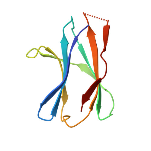 | |
UniProt | |||||
Find proteins for A0A1S4NYE1 (Paenibacillus barengoltzii) Explore A0A1S4NYE1 Go to UniProtKB: A0A1S4NYE1 | |||||
Entity Groups | |||||
| Sequence Clusters | 30% Identity50% Identity70% Identity90% Identity95% Identity100% Identity | ||||
| UniProt Group | A0A1S4NYE1 | ||||
Sequence AnnotationsExpand | |||||
| |||||
| Length ( Å ) | Angle ( ˚ ) |
|---|---|
| a = 67.424 | α = 90 |
| b = 67.424 | β = 90 |
| c = 74.988 | γ = 90 |
| Software Name | Purpose |
|---|---|
| PHENIX | refinement |
| HKL-3000 | data reduction |
| HKL-3000 | data scaling |
| PHENIX | phasing |
| Funding Organization | Location | Grant Number |
|---|---|---|
| National Natural Science Foundation of China | China | 31701537 |
RCSB PDB (citation) is hosted by
RCSB PDB is a member of the

