6A22

Ternary complex of Human ROR gamma Ligand Binding Domain With Compound T.


Experimental Data Snapshot

  • Method: X-RAY DIFFRACTION
  • Resolution: 2.55 Å
  • R-Value Free: 0.243 
  • R-Value Work: 0.217 
  • R-Value Observed: 0.219 

wwPDB Validation   3D Report Full Report


Ligand Structure Quality Assessment 


This is version 1.1 of the entry. See complete history


Literature

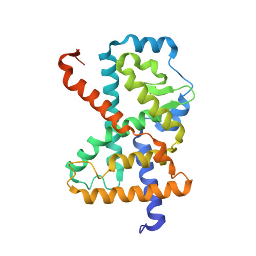
Ternary crystal structure of human ROR gamma ligand-binding-domain, an inhibitor and corepressor peptide provides a new insight into corepressor interaction

Noguchi, M.Nomura, A.Doi, S.Yamaguchi, K.Hirata, K.Shiozaki, M.Maeda, K.Hirashima, S.Kotoku, M.Yamaguchi, T.Katsuda, Y.Crowe, P.Tao, H.Thacher, S.Adachi, T.

(2018) Sci Rep 8: 17374-17374

  • DOI: https://doi.org/10.1038/s41598-018-35783-9
  • Primary Citation of Related Structures:  
    6A22

  • PubMed Abstract: 

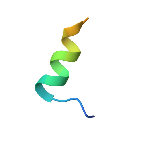
    Retinoic acid-related orphan receptor gamma (RORγ) plays pivotal roles in autoimmune diseases by controlling the lineage of interleukin 17 (IL-17)-producing CD4 + T cells (Th17 cells). Structure-based drug design has proven fruitful in the development of inhibitors targeting the ligand binding domain (LBD) of RORγ. Here, we present the crystal structure of a novel RORγ inhibitor co-complex, in the presence of a corepressor (CoR) peptide. This ternary complex with compound T reveals the structural basis for an inhibitory mechanism different from the previously reported inverse agonist. Compared to the inverse agonist, compound T induces about 2 Å shift of helix 5 (H5) backbone and side-chain conformational changes of Met365 on H5. These conformational changes correlate to reduced CoR peptide binding to RORγ-LBD in the presence of compound T, which suggests that the shift of H5 is responsible. This crystal structure analysis will provide useful information for the development of novel and efficacious drugs for autoimmune disorders.


  • Organizational Affiliation

    Pharmaceutical Frontier Research Laboratories, Central Pharmaceutical Research Institute, Japan Tobacco Inc., 1-13-2, Fukuura, Kanazawa-Ku, Yokohama, Kanagawa, 236-0004, Japan. masato.noguchi@jt.com.


Macromolecules
Find similar proteins by:  (by identity cutoff)  |  3D Structure
Entity ID: 1
MoleculeChains Sequence LengthOrganismDetailsImage
Nuclear receptor ROR-gamma
A, C, E, G
258Homo sapiensMutation(s): 2 
Gene Names: RORCNR1F3RORGRZRG
UniProt & NIH Common Fund Data Resources
Find proteins for P51449 (Homo sapiens)
Explore P51449 
Go to UniProtKB:  P51449
PHAROS:  P51449
GTEx:  ENSG00000143365 
Entity Groups  
Sequence Clusters30% Identity50% Identity70% Identity90% Identity95% Identity100% Identity
UniProt GroupP51449
Sequence Annotations
Expand
  • Reference Sequence

Find similar proteins by:  Sequence   |   3D Structure  

Entity ID: 2
MoleculeChains Sequence LengthOrganismDetailsImage
Nuclear receptor corepressor 2
B, D, F, H
22Homo sapiensMutation(s): 0 
Gene Names: NCOR2CTG26
UniProt & NIH Common Fund Data Resources
Find proteins for Q9Y618 (Homo sapiens)
Explore Q9Y618 
Go to UniProtKB:  Q9Y618
PHAROS:  Q9Y618
GTEx:  ENSG00000196498 
Entity Groups  
Sequence Clusters30% Identity50% Identity70% Identity90% Identity95% Identity100% Identity
UniProt GroupQ9Y618
Sequence Annotations
Expand
  • Reference Sequence
Experimental Data & Validation

Experimental Data

  • Method: X-RAY DIFFRACTION
  • Resolution: 2.55 Å
  • R-Value Free: 0.243 
  • R-Value Work: 0.217 
  • R-Value Observed: 0.219 
  • Space Group: P 1 21 1
Unit Cell:
Length ( Å )Angle ( ˚ )
a = 77.926α = 90
b = 73.277β = 89.99
c = 100.204γ = 90
Software Package:
Software NamePurpose
REFMACrefinement
XDSdata reduction
SCALAdata scaling
PHASERphasing

Structure Validation

View Full Validation Report


Ligand Structure Quality Assessment 


Entry History 

Deposition Data

Revision History  (Full details and data files)

  • Version 1.0: 2018-12-12
    Type: Initial release
  • Version 1.1: 2023-11-22
    Changes: Data collection, Database references, Refinement description