5ZQM

Crystal structure of human katanin AAA ATPase domain complex with ATPgammaS


Experimental Data Snapshot

  • Method: X-RAY DIFFRACTION
  • Resolution: 2.90 Å
  • R-Value Free: 0.285 
  • R-Value Work: 0.247 
  • R-Value Observed: 0.249 

wwPDB Validation   3D Report Full Report


Ligand Structure Quality Assessment 


This is version 1.1 of the entry. See complete history


Literature

Structural and Molecular Basis for Katanin-Mediated Severing of Glutamylated Microtubules.

Shin, S.C.Im, S.K.Jang, E.H.Jin, K.S.Hur, E.M.Kim, E.E.

(2019) Cell Rep 26: 1357-1367.e5

  • DOI: https://doi.org/10.1016/j.celrep.2019.01.020
  • Primary Citation of Related Structures:  
    5ZQL, 5ZQM

  • PubMed Abstract: 

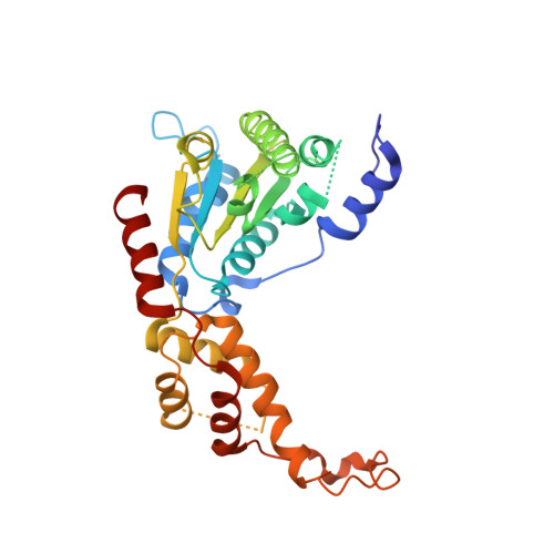
    Katanin was the first microtubule (MT)-severing enzyme discovered, but how katanin executes MT severing remains poorly understood. Here, we report X-ray crystal structures of the apo and ATPγS-bound states of the catalytic AAA domain of human katanin p60 at 3.0 and 2.9 Å resolution, respectively. Comparison of the two structures reveals conformational changes induced by ATP binding and how such changes ensure hexamer stability. Moreover, we uncover structural details of pore loops (PLs) and show that Arg283, a residue unique to katanin among MT-severing enzymes, protrudes from PL1 and lines the entry of the catalytic pore. Functional studies suggest that PL1 and Arg283 play essential roles in the recognition and remodeling of the glutamylated, C-terminal tubulin tail and regulation of axon growth. In addition, domain-swapping experiments in katanin and spastin suggest that the non-homologous N-terminal region, which contains the MT-interacting and trafficking domain and a linker, confers specificity to the severing process.


  • Organizational Affiliation

    Biomedical Research Institute, Korea Institute of Science and Technology, Seoul, 02792, Republic of Korea.


Macromolecules
Find similar proteins by:  (by identity cutoff)  |  3D Structure
Entity ID: 1
MoleculeChains Sequence LengthOrganismDetailsImage
Katanin p60 ATPase-containing subunit A1309Homo sapiensMutation(s): 0 
Gene Names: KATNA1
EC: 3.6.4.3
UniProt & NIH Common Fund Data Resources
Find proteins for O75449 (Homo sapiens)
Explore O75449 
Go to UniProtKB:  O75449
PHAROS:  O75449
GTEx:  ENSG00000186625 
Entity Groups  
Sequence Clusters30% Identity50% Identity70% Identity90% Identity95% Identity100% Identity
UniProt GroupO75449
Sequence Annotations
Expand
  • Reference Sequence
Small Molecules
Ligands 1 Unique
IDChains Name / Formula / InChI Key2D Diagram3D Interactions
AGS
Query on AGS

Download Ideal Coordinates CCD File 
B [auth A]PHOSPHOTHIOPHOSPHORIC ACID-ADENYLATE ESTER
C10 H16 N5 O12 P3 S
NLTUCYMLOPLUHL-KQYNXXCUSA-N
Experimental Data & Validation

Experimental Data

  • Method: X-RAY DIFFRACTION
  • Resolution: 2.90 Å
  • R-Value Free: 0.285 
  • R-Value Work: 0.247 
  • R-Value Observed: 0.249 
  • Space Group: P 65
Unit Cell:
Length ( Å )Angle ( ˚ )
a = 68.907α = 90
b = 68.907β = 90
c = 138.698γ = 120
Software Package:
Software NamePurpose
REFMACrefinement
HKL-2000data scaling
PDB_EXTRACTdata extraction
HKL-2000data reduction
PHENIXphasing

Structure Validation

View Full Validation Report


Ligand Structure Quality Assessment 


Entry History & Funding Information

Deposition Data


Funding OrganizationLocationGrant Number
Korea, Republic Of--

Revision History  (Full details and data files)

  • Version 1.0: 2019-02-27
    Type: Initial release
  • Version 1.1: 2023-11-22
    Changes: Data collection, Database references, Refinement description