


Crystal Structure of Selenomethionine labelled Drep4 CIDE domain
Park, H.H.To be published.
Experimental Data Snapshot
wwPDB Validation 3D Report Full Report

Entity ID: 1 | |||||
|---|---|---|---|---|---|
| Molecule | Chains | Sequence Length | Organism | Details | Image |
| DNAation factor-related protein 4 | 100 | Drosophila melanogaster | Mutation(s): 0 Gene Names: Drep4, Drep-4, drep4, Rep4, CG9414, Dmel_CG9414 | 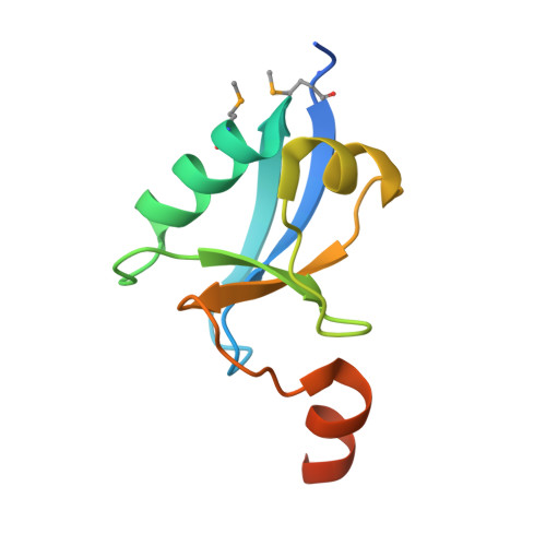 | |
UniProt | |||||
Find proteins for Q9V3H0 (Drosophila melanogaster) Explore Q9V3H0 Go to UniProtKB: Q9V3H0 | |||||
Entity Groups | |||||
| Sequence Clusters | 30% Identity50% Identity70% Identity90% Identity95% Identity100% Identity | ||||
| UniProt Group | Q9V3H0 | ||||
Sequence AnnotationsExpand | |||||
| |||||
| Modified Residues 1 Unique | |||||
|---|---|---|---|---|---|
| ID | Chains | Type | Formula | 2D Diagram | Parent |
| MSE Query on MSE | A, B, C, D, E A, B, C, D, E, F, G, H, I, J | L-PEPTIDE LINKING | C5 H11 N O2 Se |  | MET |
| Length ( Å ) | Angle ( ˚ ) |
|---|---|
| a = 56.511 | α = 102.46 |
| b = 103.722 | β = 105.81 |
| c = 103.82 | γ = 105.77 |
| Software Name | Purpose |
|---|---|
| PHENIX | refinement |
| PHENIX | data reduction |
| HKL-2000 | data scaling |
| PHENIX | phasing |
RCSB PDB (citation) is hosted by
RCSB PDB is a member of the

