5X24

Crystal structure of CYP2C9 genetic variant I359L (*3) in complex with multiple losartan molecules


Experimental Data Snapshot

  • Method: X-RAY DIFFRACTION
  • Resolution: 2.48 Å
  • R-Value Free: 0.270 
  • R-Value Work: 0.229 
  • R-Value Observed: 0.231 

wwPDB Validation   3D Report Full Report


Ligand Structure Quality Assessment 


This is version 1.2 of the entry. See complete history


Literature

Structural Basis of Single-Nucleotide Polymorphisms in Cytochrome P450 2C9

Maekawa, K.Adachi, M.Matsuzawa, Y.Zhang, Q.Kuroki, R.Saito, Y.Shah, M.B.

(2017) Biochemistry 56: 5476-5480

  • DOI: https://doi.org/10.1021/acs.biochem.7b00795
  • Primary Citation of Related Structures:  
    5X23, 5X24, 5XXI

  • PubMed Abstract: 

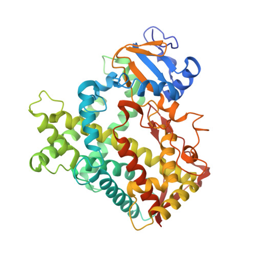
    Single-nucleotide polymorphisms in drug-metabolizing cytochrome P450 (CYP) enzymes are important contributors to interindividual differences in drug metabolism leading to adverse drug reactions. Despite their extensive characterization and importance in pharmacogenetics of clinical drugs, the structural basis of CYP polymorphisms has remained scant. Here we report the crystal structures of human CYP2C9 and its polymorphic variants, *3 (I359L) and *30 (A477T), with an antihypertensive drug losartan. The structures show distinct interaction and occupation of losartan in the active site, the access channel, and the peripheral binding site. The I359L substitution located far from the active site remarkably altered the residue side chains near the active site and the access channel, whereas the T477 substitution illustrated hydrogen-bonding interaction with the reoriented side chain of Q214. The results yield structural insights into the reduced catalytic activity of the CYP2C9 variants and have important implications for understanding genetic polymorphisms in CYP-mediated drug metabolism.


  • Organizational Affiliation

    Department of Analytical Chemistry, Faculty of Pharmaceutical Sciences, Doshisha Women's College of Liberal Arts , Kodo, Kyotanabe, Kyoto 610-0395, Japan.


Macromolecules
Find similar proteins by:  (by identity cutoff)  |  3D Structure
Entity ID: 1
MoleculeChains Sequence LengthOrganismDetailsImage
Cytochrome P450 2C9476Homo sapiensMutation(s): 2 
Gene Names: CYP2C9CYP2C10
EC: 1.14.13 (PDB Primary Data), 1.14.13.80 (PDB Primary Data), 1.14.13.48 (PDB Primary Data), 1.14.13.49 (PDB Primary Data), 1.14.99.38 (PDB Primary Data)
UniProt & NIH Common Fund Data Resources
Find proteins for P11712 (Homo sapiens)
Explore P11712 
Go to UniProtKB:  P11712
PHAROS:  P11712
GTEx:  ENSG00000138109 
Entity Groups  
Sequence Clusters30% Identity50% Identity70% Identity90% Identity95% Identity100% Identity
UniProt GroupP11712
Sequence Annotations
Expand
  • Reference Sequence
Experimental Data & Validation

Experimental Data

  • Method: X-RAY DIFFRACTION
  • Resolution: 2.48 Å
  • R-Value Free: 0.270 
  • R-Value Work: 0.229 
  • R-Value Observed: 0.231 
  • Space Group: I 2 2 2
Unit Cell:
Length ( Å )Angle ( ˚ )
a = 74.858α = 90
b = 141.777β = 90
c = 160.656γ = 90
Software Package:
Software NamePurpose
REFMACrefinement
HKL-2000data reduction
HKL-2000data scaling
PHENIXphasing

Structure Validation

View Full Validation Report


Ligand Structure Quality Assessment 


Entry History & Funding Information

Deposition Data


Funding OrganizationLocationGrant Number
JapanKAKENHI22590054
JapanKAKENHI25293128

Revision History  (Full details and data files)

  • Version 1.0: 2017-10-25
    Type: Initial release
  • Version 1.1: 2017-11-01
    Changes: Database references
  • Version 1.2: 2023-11-22
    Changes: Data collection, Database references, Refinement description