5WHT

Crystal structure of 3'SL bound PltB


Experimental Data Snapshot

  • Method: X-RAY DIFFRACTION
  • Resolution: 1.93 Å
  • R-Value Free: 0.211 
  • R-Value Work: 0.160 
  • R-Value Observed: 0.163 

wwPDB Validation   3D Report Full Report


Ligand Structure Quality Assessment 


This is version 2.1 of the entry. See complete history


Literature

Evolution of host adaptation in the Salmonella typhoid toxin.

Gao, X.Deng, L.Stack, G.Yu, H.Chen, X.Naito-Matsui, Y.Varki, A.Galan, J.E.

(2017) Nat Microbiol 2: 1592-1599

  • DOI: https://doi.org/10.1038/s41564-017-0033-2
  • Primary Citation of Related Structures:  
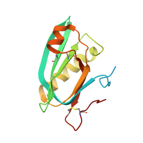
    5WHT, 5WHU, 5WHV

  • PubMed Abstract: 

    The evolution of virulence traits is central for the emergence or re-emergence of microbial pathogens and for their adaptation to a specific host 1-5 . Typhoid toxin is an essential virulence factor of the human-adapted bacterial pathogen Salmonella Typhi 6,7 , the cause of typhoid fever in humans 8-12 . Typhoid toxin has a unique A 2 B 5 architecture with two covalently linked enzymatic 'A' subunits, PltA and CdtB, associated with a homopentameric 'B' subunit made up of PltB, which has binding specificity for the N-acetylneuraminic acid (Neu5Ac) sialoglycans 6,13 prominently present in humans 14 . Here, we examine the functional and structural relationship between typhoid toxin and ArtAB, an evolutionarily related AB 5 toxin encoded by the broad-host Salmonella Typhimurium 15 . We find that ArtA and ArtB, homologues of PltA and PltB, can form a functional complex with the typhoid toxin CdtB subunit after substitution of a single amino acid in ArtA, while ArtB can form a functional complex with wild-type PltA and CdtB. We also found that, after addition of a single-terminal Cys residue, a CdtB homologue from cytolethal distending toxin can form a functional complex with ArtA and ArtB. In line with the broad host specificity of S. Typhimurium, we found that ArtB binds human glycans, terminated in N-acetylneuraminic acid, as well as glycans terminated in N-glycolylneuraminic acid (Neu5Gc), which are expressed in most other mammals 14 . The atomic structure of ArtB bound to its receptor shows the presence of an additional glycan-binding site, which broadens its binding specificity. Despite equivalent toxicity in vitro, we found that the ArtB/PltA/CdtB chimaeric toxin exhibits reduced lethality in an animal model, indicating that the host specialization of typhoid toxin has optimized its targeting mechanisms to the human host. This is a remarkable example of a toxin evolving to broaden its enzymatic activities and adapt to a specific host.


  • Organizational Affiliation

    Department of Microbial Pathogenesis, Yale University School of Medicine, New Haven, CT, 06536, USA.


Macromolecules
Find similar proteins by:  (by identity cutoff)  |  3D Structure
Entity ID: 1
MoleculeChains Sequence LengthOrganismDetailsImage
Putative pertussis-like toxin subunit
A, B, C, D, E
138Salmonella enterica subsp. enterica serovar TyphiMutation(s): 0 
Gene Names: STY1891t1107
UniProt
Find proteins for Q8Z6A3 (Salmonella typhi)
Explore Q8Z6A3 
Go to UniProtKB:  Q8Z6A3
Entity Groups  
Sequence Clusters30% Identity50% Identity70% Identity90% Identity95% Identity100% Identity
UniProt GroupQ8Z6A3
Sequence Annotations
Expand
  • Reference Sequence
Oligosaccharides

Help

Entity ID: 2
MoleculeChains Length2D Diagram Glycosylation3D Interactions
N-acetyl-alpha-neuraminic acid-(2-3)-beta-D-galactopyranose-(1-4)-alpha-D-glucopyranose
F
3N/A
Glycosylation Resources
GlyTouCan:  G92323ZH
GlyCosmos:  G92323ZH
GlyGen:  G92323ZH
Entity ID: 3
MoleculeChains Length2D Diagram Glycosylation3D Interactions
N-acetyl-alpha-neuraminic acid-(2-3)-beta-D-galactopyranose
G
2N/A
Glycosylation Resources
GlyTouCan:  G30207PZ
GlyCosmos:  G30207PZ
GlyGen:  G30207PZ
Small Molecules
Ligands 3 Unique
IDChains Name / Formula / InChI Key2D Diagram3D Interactions
SIA
Query on SIA

Download Ideal Coordinates CCD File 
I [auth B],
O [auth D]
N-acetyl-alpha-neuraminic acid
C11 H19 N O9
SQVRNKJHWKZAKO-YRMXFSIDSA-N
PEG
Query on PEG

Download Ideal Coordinates CCD File 
M [auth B],
R [auth D],
T [auth E],
U [auth E],
V [auth E]
DI(HYDROXYETHYL)ETHER
C4 H10 O3
MTHSVFCYNBDYFN-UHFFFAOYSA-N
ACT
Query on ACT

Download Ideal Coordinates CCD File 
H [auth A]
J [auth B]
K [auth B]
L [auth B]
N [auth C]
H [auth A],
J [auth B],
K [auth B],
L [auth B],
N [auth C],
P [auth D],
Q [auth D],
S [auth E]
ACETATE ION
C2 H3 O2
QTBSBXVTEAMEQO-UHFFFAOYSA-M
Biologically Interesting Molecules (External Reference) 1 Unique
Experimental Data & Validation

Experimental Data

  • Method: X-RAY DIFFRACTION
  • Resolution: 1.93 Å
  • R-Value Free: 0.211 
  • R-Value Work: 0.160 
  • R-Value Observed: 0.163 
  • Space Group: P 21 21 21
Unit Cell:
Length ( Å )Angle ( ˚ )
a = 60.379α = 90
b = 95.682β = 90
c = 117.173γ = 90
Software Package:
Software NamePurpose
PHENIXrefinement
HKL-2000data collection
PHASERphasing

Structure Validation

View Full Validation Report


Ligand Structure Quality Assessment 


Entry History & Funding Information

Deposition Data


Funding OrganizationLocationGrant Number
National Institutes of Health/National Institute Of Allergy and Infectious Diseases (NIH/NIAID)United StatesAI079022

Revision History  (Full details and data files)

  • Version 1.0: 2017-10-25
    Type: Initial release
  • Version 1.1: 2017-12-06
    Changes: Database references
  • Version 1.2: 2019-12-11
    Changes: Author supporting evidence, Data collection
  • Version 2.0: 2020-07-29
    Type: Remediation
    Reason: Carbohydrate remediation
    Changes: Atomic model, Data collection, Derived calculations, Structure summary
  • Version 2.1: 2023-10-04
    Changes: Data collection, Database references, Derived calculations, Refinement description, Structure summary