5VR0

Crystal structure of glucose isomerase from Streptomyces rubiginosus


Experimental Data Snapshot

  • Method: X-RAY DIFFRACTION
  • Resolution: 1.70 Å
  • R-Value Free: 0.142 
  • R-Value Work: 0.119 
  • R-Value Observed: 0.120 

wwPDB Validation   3D Report Full Report


This is version 1.3 of the entry. See complete history


Literature

Real-space analysis of radiation-induced specific changes with independent component analysis.

Borek, D.Bromberg, R.Hattne, J.Otwinowski, Z.

(2018) J Synchrotron Radiat 25: 451-467

  • DOI: https://doi.org/10.1107/S1600577517018148
  • Primary Citation of Related Structures:  
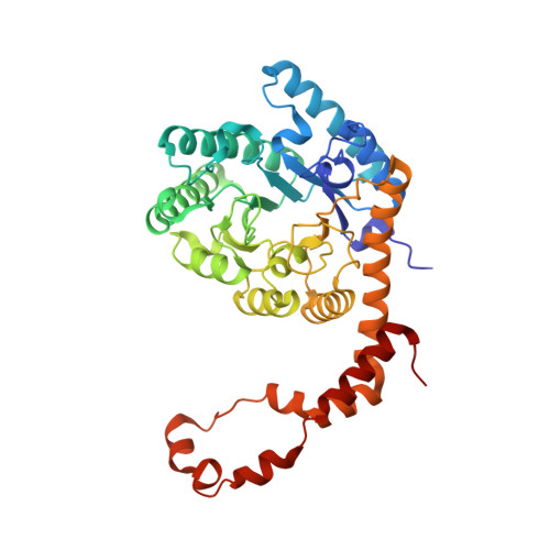
    5VR0

  • PubMed Abstract: 

    A method of analysis is presented that allows for the separation of specific radiation-induced changes into distinct components in real space. The method relies on independent component analysis (ICA) and can be effectively applied to electron density maps and other types of maps, provided that they can be represented as sets of numbers on a grid. Here, for glucose isomerase crystals, ICA was used in a proof-of-concept analysis to separate temperature-dependent and temperature-independent components of specific radiation-induced changes for data sets acquired from multiple crystals across multiple temperatures. ICA identified two components, with the temperature-independent component being responsible for the majority of specific radiation-induced changes at temperatures below 130 K. The patterns of specific temperature-independent radiation-induced changes suggest a contribution from the tunnelling of electron holes as a possible explanation. In the second case, where a group of 22 data sets was collected on a single thaumatin crystal, ICA was used in another type of analysis to separate specific radiation-induced effects happening on different exposure-level scales. Here, ICA identified two components of specific radiation-induced changes that likely result from radiation-induced chemical reactions progressing with different rates at different locations in the structure. In addition, ICA unexpectedly identified the radiation-damage state corresponding to reduced disulfide bridges rather than the zero-dose extrapolated state as the highest contrast structure. The application of ICA to the analysis of specific radiation-induced changes in real space and the data pre-processing for ICA that relies on singular value decomposition, which was used previously in data space to validate a two-component physical model of X-ray radiation-induced changes, are discussed in detail. This work lays a foundation for a better understanding of protein-specific radiation chemistries and provides a framework for analysing effects of specific radiation damage in crystallographic and cryo-EM experiments.


  • Organizational Affiliation

    Department of Biophysics, University of Texas Southwestern Medical Center, 5323 Harry Hines Blvd, Dallas, TX 75390, USA.


Macromolecules
Find similar proteins by:  (by identity cutoff)  |  3D Structure
Entity ID: 1
MoleculeChains Sequence LengthOrganismDetailsImage
Xylose isomerase388Streptomyces rubiginosusMutation(s): 0 
Gene Names: xylA
EC: 5.3.1.5
UniProt
Find proteins for P24300 (Streptomyces rubiginosus)
Explore P24300 
Go to UniProtKB:  P24300
Entity Groups  
Sequence Clusters30% Identity50% Identity70% Identity90% Identity95% Identity100% Identity
UniProt GroupP24300
Sequence Annotations
Expand
  • Reference Sequence
Small Molecules
Ligands 4 Unique
IDChains Name / Formula / InChI Key2D Diagram3D Interactions
MRD
Query on MRD

Download Ideal Coordinates CCD File 
B [auth A](4R)-2-METHYLPENTANE-2,4-DIOL
C6 H14 O2
SVTBMSDMJJWYQN-RXMQYKEDSA-N
MN
Query on MN

Download Ideal Coordinates CCD File 
C [auth A],
D [auth A]
MANGANESE (II) ION
Mn
WAEMQWOKJMHJLA-UHFFFAOYSA-N
CA
Query on CA

Download Ideal Coordinates CCD File 
Q [auth A],
R [auth A],
S [auth A]
CALCIUM ION
Ca
BHPQYMZQTOCNFJ-UHFFFAOYSA-N
CL
Query on CL

Download Ideal Coordinates CCD File 
E [auth A]
F [auth A]
G [auth A]
H [auth A]
I [auth A]
E [auth A],
F [auth A],
G [auth A],
H [auth A],
I [auth A],
J [auth A],
K [auth A],
L [auth A],
M [auth A],
N [auth A],
O [auth A],
P [auth A]
CHLORIDE ION
Cl
VEXZGXHMUGYJMC-UHFFFAOYSA-M
Experimental Data & Validation

Experimental Data

  • Method: X-RAY DIFFRACTION
  • Resolution: 1.70 Å
  • R-Value Free: 0.142 
  • R-Value Work: 0.119 
  • R-Value Observed: 0.120 
  • Space Group: I 2 2 2
Unit Cell:
Length ( Å )Angle ( ˚ )
a = 92.854α = 90
b = 98.061β = 90
c = 102.279γ = 90
Software Package:
Software NamePurpose
REFMACrefinement
HKL-2000data reduction
HKL-3000data scaling
REFMACphasing

Structure Validation

View Full Validation Report


Entry History & Funding Information

Deposition Data


Funding OrganizationLocationGrant Number
National Institutes of Health/National Institute of General Medical Sciences (NIH/NIGMS)United StatesR01GM118619
National Institutes of Health/National Institute of General Medical Sciences (NIH/NIGMS)United StatesR01GM117080
National Institutes of Health/National Institute of General Medical Sciences (NIH/NIGMS)United StatesR01GM053163

Revision History  (Full details and data files)

  • Version 1.0: 2018-05-02
    Type: Initial release
  • Version 1.1: 2019-02-20
    Changes: Author supporting evidence, Data collection
  • Version 1.2: 2020-01-01
    Changes: Author supporting evidence
  • Version 1.3: 2023-10-04
    Changes: Data collection, Database references, Derived calculations, Refinement description