


Crystal structure of nucleoside diphosphate kinase from Neisseria gonorrhoeae in complex with citrate
Abendroth, J., Mayclin, S.J., Lorimer, D.D., Edwards, T.E.To be published.
Experimental Data Snapshot
wwPDB Validation 3D Report Full Report

Entity ID: 1 | |||||
|---|---|---|---|---|---|
| Molecule | Chains | Sequence Length | Organism | Details | Image |
| Nucleoside diphosphate kinase | 149 | Neisseria gonorrhoeae NCCP11945 | Mutation(s): 0 Gene Names: ndk, NGK_1320 EC: 2.7.4.6 | 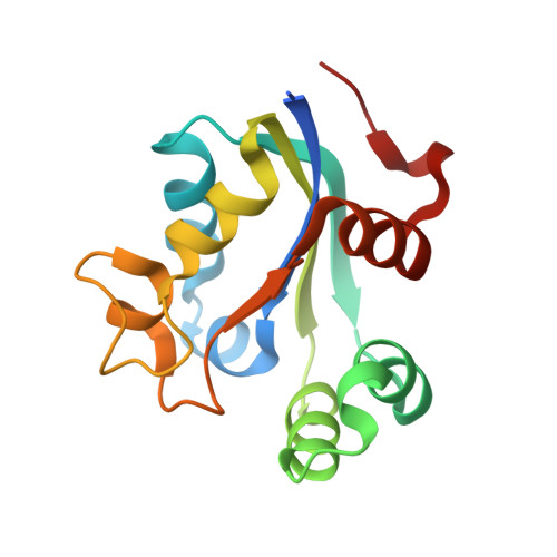 | |
UniProt | |||||
Find proteins for B4RMG0 (Neisseria gonorrhoeae (strain NCCP11945)) Explore B4RMG0 Go to UniProtKB: B4RMG0 | |||||
Entity Groups | |||||
| Sequence Clusters | 30% Identity50% Identity70% Identity90% Identity95% Identity100% Identity | ||||
| UniProt Group | B4RMG0 | ||||
Sequence AnnotationsExpand | |||||
| |||||
| Ligands 2 Unique | |||||
|---|---|---|---|---|---|
| ID | Chains | Name / Formula / InChI Key | 2D Diagram | 3D Interactions | |
| CIT Query on CIT | AA [auth E] AB [auth P] CA [auth F] EA [auth G] HA [auth H] | CITRIC ACID C6 H8 O7 KRKNYBCHXYNGOX-UHFFFAOYSA-N |  | ||
| EDO Query on EDO | BA [auth E] BB [auth P] DA [auth F] FA [auth G] GA [auth H] | 1,2-ETHANEDIOL C2 H6 O2 LYCAIKOWRPUZTN-UHFFFAOYSA-N |  | ||
| Length ( Å ) | Angle ( ˚ ) |
|---|---|
| a = 57.24 | α = 104.31 |
| b = 112.35 | β = 98.5 |
| c = 112.87 | γ = 105.39 |
| Software Name | Purpose |
|---|---|
| PHENIX | refinement |
| XSCALE | data scaling |
| PDB_EXTRACT | data extraction |
| XDS | data reduction |
| MOLREP | phasing |
RCSB PDB (citation) is hosted by
RCSB PDB is a member of the

