5V5Z

Structure of CYP51 from the pathogen Candida albicans


Experimental Data Snapshot

  • Method: X-RAY DIFFRACTION
  • Resolution: 2.90 Å
  • R-Value Free: 0.269 
  • R-Value Work: 0.219 
  • R-Value Observed: 0.221 

wwPDB Validation   3D Report Full Report


Ligand Structure Quality Assessment 


This is version 1.5 of the entry. See complete history


Literature

Crystal Structures of Full-Length Lanosterol 14 alpha-Demethylases of Prominent Fungal Pathogens Candida albicans and Candida glabrata Provide Tools for Antifungal Discovery.

Keniya, M.V.Sabherwal, M.Wilson, R.K.Woods, M.A.Sagatova, A.A.Tyndall, J.D.A.Monk, B.C.

(2018) Antimicrob Agents Chemother 62

  • DOI: https://doi.org/10.1128/AAC.01134-18
  • Primary Citation of Related Structures:  
    5JLC, 5V5Z

  • PubMed Abstract: 

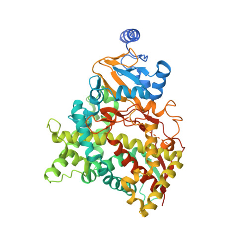
    Targeting lanosterol 14α-demethylase (LDM) with azole drugs provides prophylaxis and treatments for superficial and disseminated fungal infections, but cure rates are not optimal for immunocompromised patients and individuals with comorbidities. The efficacy of azole drugs has also been reduced due to the emergence of drug-resistant fungal pathogens. We have addressed the need to improve the potency, spectrum, and specificity for azoles by expressing in Saccharomyces cerevisiae functional, recombinant, hexahistidine-tagged, full-length Candida albicans LDM (CaLDM6×His) and Candida glabrata LDM (CgLDM6×His) and determining their X-ray crystal structures. The crystal structures of CaLDM6×His, CgLDM6×His, and ScLDM6×His have the same fold and bind itraconazole in nearly identical conformations. The catalytic domains of the full-length LDMs have the same fold as the CaLDM6×His catalytic domain in complex with posaconazole, with minor structural differences within the ligand binding pocket. Our structures give insight into the LDM reaction mechanism and phenotypes of single-site CaLDM mutations. This study provides a practical basis for the structure-directed discovery of novel antifungals that target LDMs of fungal pathogens.


  • Organizational Affiliation

    Sir John Walsh Research Institute, Faculty of Dentistry, University of Otago, Dunedin, New Zealand.


Macromolecules
Find similar proteins by:  (by identity cutoff)  |  3D Structure
Entity ID: 1
MoleculeChains Sequence LengthOrganismDetailsImage
Lanosterol 14-alpha demethylase537Candida albicans SC5314Mutation(s): 0 
Gene Names: ERG11CYP51ERG16CAALFM_C500660CACaO19.922
EC: 1.14.13.70
Membrane Entity: Yes 
UniProt
Find proteins for P10613 (Candida albicans (strain SC5314 / ATCC MYA-2876))
Explore P10613 
Go to UniProtKB:  P10613
Entity Groups  
Sequence Clusters30% Identity50% Identity70% Identity90% Identity95% Identity100% Identity
UniProt GroupP10613
Sequence Annotations
Expand
  • Reference Sequence
Small Molecules
Ligands 2 Unique
IDChains Name / Formula / InChI Key2D Diagram3D Interactions
1YN
Query on 1YN

Download Ideal Coordinates CCD File 
C [auth A]2-[(2R)-butan-2-yl]-4-{4-[4-(4-{[(2R,4S)-2-(2,4-dichlorophenyl)-2-(1H-1,2,4-triazol-1-ylmethyl)-1,3-dioxolan-4-yl]methoxy}phenyl)piperazin-1-yl]phenyl}-2,4-dihydro-3H-1,2,4-triazol-3-one
C35 H38 Cl2 N8 O4
VHVPQPYKVGDNFY-DFMJLFEVSA-N
HEM
Query on HEM

Download Ideal Coordinates CCD File 
B [auth A]PROTOPORPHYRIN IX CONTAINING FE
C34 H32 Fe N4 O4
KABFMIBPWCXCRK-RGGAHWMASA-L
Experimental Data & Validation

Experimental Data

  • Method: X-RAY DIFFRACTION
  • Resolution: 2.90 Å
  • R-Value Free: 0.269 
  • R-Value Work: 0.219 
  • R-Value Observed: 0.221 
  • Space Group: P 2 21 21
Unit Cell:
Length ( Å )Angle ( ˚ )
a = 91.94α = 90
b = 92.51β = 90
c = 101.53γ = 90
Software Package:
Software NamePurpose
PHENIXrefinement
MOSFLMdata reduction
Aimlessdata scaling
PHASERphasing

Structure Validation

View Full Validation Report


Ligand Structure Quality Assessment 


Entry History & Funding Information

Deposition Data


Funding OrganizationLocationGrant Number
Health Research Council (HRC)New Zealand--
Marsden FundNew Zealand--

Revision History  (Full details and data files)

  • Version 1.0: 2017-03-29
    Type: Initial release
  • Version 1.1: 2017-04-12
    Changes: Structure summary
  • Version 1.2: 2017-09-20
    Changes: Author supporting evidence, Data collection
  • Version 1.3: 2020-01-01
    Changes: Author supporting evidence
  • Version 1.4: 2020-02-12
    Changes: Database references
  • Version 1.5: 2023-10-04
    Changes: Data collection, Database references, Refinement description