5URY

Crystal structure of Frizzled 5 CRD in complex with PAM


Experimental Data Snapshot

  • Method: X-RAY DIFFRACTION
  • Resolution: 2.10 Å
  • R-Value Free: 0.206 
  • R-Value Work: 0.170 
  • R-Value Observed: 0.172 

wwPDB Validation   3D Report Full Report


Ligand Structure Quality Assessment 


This is version 2.0 of the entry. See complete history


Literature

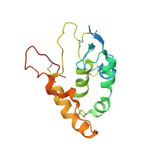
Unsaturated fatty acyl recognition by Frizzled receptors mediates dimerization upon Wnt ligand binding.

Nile, A.H.Mukund, S.Stanger, K.Wang, W.Hannoush, R.N.

(2017) Proc Natl Acad Sci U S A 114: 4147-4152

  • DOI: https://doi.org/10.1073/pnas.1618293114
  • Primary Citation of Related Structures:  
    5T44, 5URV, 5URY, 5URZ

  • PubMed Abstract: 

    Frizzled (FZD) receptors mediate Wnt signaling in diverse processes ranging from bone growth to stem cell activity. Moreover, high FZD receptor expression at the cell surface contributes to overactive Wnt signaling in subsets of pancreatic, ovarian, gastric, and colorectal tumors. Despite the progress in biochemical understanding of Wnt-FZD receptor interactions, the molecular basis for recognition of Wnt cis -unsaturated fatty acyl groups by the cysteine-rich domain (CRD) of FZD receptors remains elusive. Here, we determined a crystal structure of human FZD7 CRD unexpectedly bound to a 24-carbon fatty acid. We also report a crystal structure of human FZD5 CRD bound to C16:1 cis -Δ9 unsaturated fatty acid. Both structures reveal a dimeric arrangement of the CRD. The lipid-binding groove exhibits flexibility and spans both monomers, adopting a U-shaped geometry that accommodates the fatty acid. Re-evaluation of the published mouse FZD8 CRD structure reveals that it also shares the same architecture as FZD5 and FZD7 CRDs. Our results define a common molecular mechanism for recognition of the cis -unsaturated fatty acyl group, a necessary posttranslational modification of Wnts, by multiple FZD receptors. The fatty acid bridges two CRD monomers, implying that Wnt binding mediates FZD receptor dimerization. Our data uncover possibilities for the arrangement of Wnt-FZD CRD complexes and shed structural insights that could aide in the identification of pharmacological strategies to modulate FZD receptor function.


  • Organizational Affiliation

    Department of Early Discovery Biochemistry, Genentech, South San Francisco, CA 94080.


Macromolecules
Find similar proteins by:  (by identity cutoff)  |  3D Structure
Entity ID: 1
MoleculeChains Sequence LengthOrganismDetailsImage
Frizzled-5
A, B
146Homo sapiensMutation(s): 0 
Gene Names: FZD5C2orf31
UniProt & NIH Common Fund Data Resources
Find proteins for Q13467 (Homo sapiens)
Explore Q13467 
Go to UniProtKB:  Q13467
PHAROS:  Q13467
GTEx:  ENSG00000163251 
Entity Groups  
Sequence Clusters30% Identity50% Identity70% Identity90% Identity95% Identity100% Identity
UniProt GroupQ13467
Sequence Annotations
Expand
  • Reference Sequence
Oligosaccharides

Help

Entity ID: 2
MoleculeChains Length2D Diagram Glycosylation3D Interactions
alpha-L-fucopyranose-(1-3)-[alpha-L-fucopyranose-(1-4)][alpha-L-fucopyranose-(1-6)]2-acetamido-2-deoxy-beta-D-glucopyranose
C
4N-Glycosylation
Glycosylation Resources
GlyTouCan:  G47513BB
GlyCosmos:  G47513BB
Experimental Data & Validation

Experimental Data

  • Method: X-RAY DIFFRACTION
  • Resolution: 2.10 Å
  • R-Value Free: 0.206 
  • R-Value Work: 0.170 
  • R-Value Observed: 0.172 
  • Space Group: P 31 2 1
Unit Cell:
Length ( Å )Angle ( ˚ )
a = 123.412α = 90
b = 123.412β = 90
c = 46.951γ = 120
Software Package:
Software NamePurpose
PHENIXrefinement
DENZOdata reduction
SCALEPACKdata scaling
PHASERphasing

Structure Validation

View Full Validation Report


Ligand Structure Quality Assessment 


Entry History 

Deposition Data

Revision History  (Full details and data files)

  • Version 1.0: 2017-05-10
    Type: Initial release
  • Version 2.0: 2020-07-29
    Type: Remediation
    Reason: Carbohydrate remediation
    Changes: Atomic model, Data collection, Derived calculations, Structure summary