5UMO

STRUCTURE OF EXTRACELLULAR SIGNAL-REGULATED KINASE


Experimental Data Snapshot

  • Method: X-RAY DIFFRACTION
  • Resolution: 2.26 Å
  • R-Value Free: 0.202 
  • R-Value Work: 0.147 
  • R-Value Observed: 0.150 

wwPDB Validation   3D Report Full Report


This is version 1.4 of the entry. See complete history


Literature

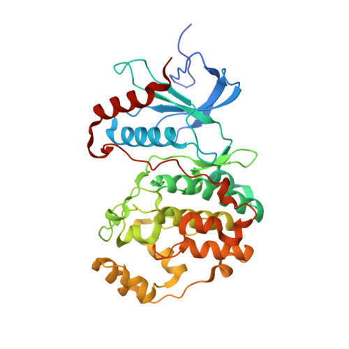
Atomic structure of the MAP kinase ERK2 at 2.3 A resolution.

Zhang, F.Strand, A.Robbins, D.Cobb, M.H.Goldsmith, E.J.

(1994) Nature 367: 704-711

  • DOI: https://doi.org/10.1038/367704a0
  • Primary Citation of Related Structures:  
    5UMO

  • PubMed Abstract: 

    The structure of the MAP kinase ERK2, a ubiquitous protein kinase target for regulation by Ras and Raf, has been solved in its unphosphorylated low-activity conformation to a resolution of 2.3 A. The two domains of unphosphorylated ERK2 are farther apart than in the active conformation of cAMP-dependent protein kinase and the peptide-binding site is blocked by tyrosine 185, one of the two residues that are phosphorylated in the active enzyme. Activation of ERK2 is thus likely to involve both global and local conformational changes.


  • Organizational Affiliation

    Department of Biochemistry, University of Texas Southwestern Medical Center at Dallas.


Macromolecules
Find similar proteins by:  (by identity cutoff)  |  3D Structure
Entity ID: 1
MoleculeChains Sequence LengthOrganismDetailsImage
Mitogen-activated protein kinase 1364Rattus norvegicusMutation(s): 0 
Gene Names: Mapk1Erk2MapkPrkm1
EC: 2.7.11.24
UniProt
Find proteins for P63086 (Rattus norvegicus)
Explore P63086 
Go to UniProtKB:  P63086
Entity Groups  
Sequence Clusters30% Identity50% Identity70% Identity90% Identity95% Identity100% Identity
UniProt GroupP63086
Sequence Annotations
Expand
  • Reference Sequence
Small Molecules
Ligands 1 Unique
IDChains Name / Formula / InChI Key2D Diagram3D Interactions
SO4
Query on SO4

Download Ideal Coordinates CCD File 
B [auth A]SULFATE ION
O4 S
QAOWNCQODCNURD-UHFFFAOYSA-L
Experimental Data & Validation

Experimental Data

  • Method: X-RAY DIFFRACTION
  • Resolution: 2.26 Å
  • R-Value Free: 0.202 
  • R-Value Work: 0.147 
  • R-Value Observed: 0.150 
  • Space Group: P 1 21 1
Unit Cell:
Length ( Å )Angle ( ˚ )
a = 49.32α = 90
b = 72.42β = 109.5
c = 61.25γ = 90
Software Package:
Software NamePurpose
HKL-2000data collection
SCALEPACKdata scaling
MOLREPphasing
REFMACrefinement
PDB_EXTRACTdata extraction
HKL-2000data reduction
Sir2014phasing
DENZOdata reduction

Structure Validation

View Full Validation Report


Entry History 

Deposition Data

Revision History  (Full details and data files)

  • Version 1.0: 2017-03-01
    Type: Initial release
  • Version 1.1: 2017-09-27
    Changes: Author supporting evidence, Refinement description
  • Version 1.2: 2017-11-22
    Changes: Advisory
  • Version 1.3: 2018-03-07
    Changes: Data collection
  • Version 1.4: 2022-04-13
    Changes: Database references