5U85

Murine saposin-D (SapD), open conformation


Experimental Data Snapshot

  • Method: X-RAY DIFFRACTION
  • Resolution: 1.65 Å
  • R-Value Free: 0.186 
  • R-Value Work: 0.166 
  • R-Value Observed: 0.167 

wwPDB Validation   3D Report Full Report


This is version 1.3 of the entry. See complete history


Literature

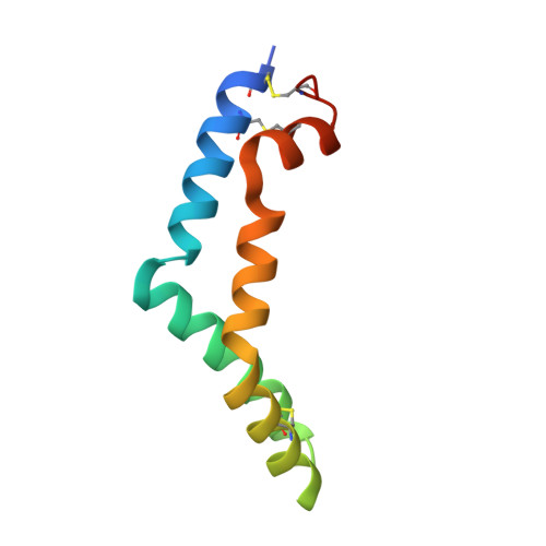
Crystal structure of saposin D in an open conformation.

Gebai, A.Gorelik, A.Nagar, B.

(2018) J Struct Biol 204: 145-150

  • DOI: https://doi.org/10.1016/j.jsb.2018.07.011
  • Primary Citation of Related Structures:  
    5U85

  • PubMed Abstract: 

    Saposins are accessory proteins that aid in the degradation of sphingolipids by hydrolytic enzymes. Their structure usually comprises four α-helices arranged in various conformations including an open, V-shaped form that is generally associated with the ability to interact with membranes and/or enzymes to accentuate activity. Saposin D is required by the lysosomal hydrolase, acid ceramidase, which breaks down ceramide into sphingosine and free fatty acid, to display optimal activity. The structure of saposin D was previously determined in an inactive conformation, revealing a monomeric, closed and compact form. Here, we present the crystal structure of the open, V-shaped form of saposin D. The overall shape is similar to the open conformation found in other saposins with slight differences in the angles between the α-helices. The structure forms a dimer that serves to stabilize the hydrophobic surface exposed in the open form, which results in an internal, hydrophobic cavity that could be used to carry extracted membrane lipids.


  • Organizational Affiliation

    Department of Biochemistry and Groupe de Recherche Axé sur la Structure des Protéines, McGill University, Montreal, QC H3G 0B1, Canada.


Macromolecules
Find similar proteins by:  (by identity cutoff)  |  3D Structure
Entity ID: 1
MoleculeChains Sequence LengthOrganismDetailsImage
Saposin-D
A, B
83Mus musculusMutation(s): 0 
Gene Names: PsapSgp1
UniProt
Find proteins for Q61207 (Mus musculus)
Explore Q61207 
Go to UniProtKB:  Q61207
Entity Groups  
Sequence Clusters30% Identity50% Identity70% Identity90% Identity95% Identity100% Identity
UniProt GroupQ61207
Sequence Annotations
Expand
  • Reference Sequence
Experimental Data & Validation

Experimental Data

  • Method: X-RAY DIFFRACTION
  • Resolution: 1.65 Å
  • R-Value Free: 0.186 
  • R-Value Work: 0.166 
  • R-Value Observed: 0.167 
  • Space Group: P 21 21 21
Unit Cell:
Length ( Å )Angle ( ˚ )
a = 41.65α = 90
b = 43.163β = 90
c = 88.822γ = 90
Software Package:
Software NamePurpose
PHENIXrefinement
HKL-2000data reduction
HKL-2000data scaling
ACORNphasing

Structure Validation

View Full Validation Report


Entry History 

Deposition Data

Revision History  (Full details and data files)

  • Version 1.0: 2018-06-13
    Type: Initial release
  • Version 1.1: 2018-08-01
    Changes: Data collection, Database references
  • Version 1.2: 2018-10-10
    Changes: Data collection, Database references, Structure summary
  • Version 1.3: 2019-05-22
    Changes: Data collection, Structure summary