5U1T

Crystal structure of the Saccharomyces cerevisiae separase-securin complex at 2.6 angstrom resolution

  • Classification: HYDROLASE
  • Organism(s): Saccharomyces cerevisiae S288C
  • Expression System: Trichoplusia ni
  • Mutation(s): No 

  • Deposited: 2016-11-29 Released: 2017-02-08 
  • Deposition Author(s): Luo, S., Tong, L.
  • Funding Organization(s): National Institutes of Health/National Institute of General Medical Sciences (NIH/NIGMS), National Institutes of Health/Office of the Director

Experimental Data Snapshot

  • Method: X-RAY DIFFRACTION
  • Resolution: 2.60 Å
  • R-Value Free: 0.264 
  • R-Value Work: 0.221 
  • R-Value Observed: 0.222 

wwPDB Validation   3D Report Full Report


This is version 1.5 of the entry. See complete history


Literature

Molecular mechanism for the regulation of yeast separase by securin.

Luo, S.Tong, L.

(2017) Nature 542: 255-259

  • DOI: https://doi.org/10.1038/nature21061
  • Primary Citation of Related Structures:  
    5U1S, 5U1T

  • PubMed Abstract: 

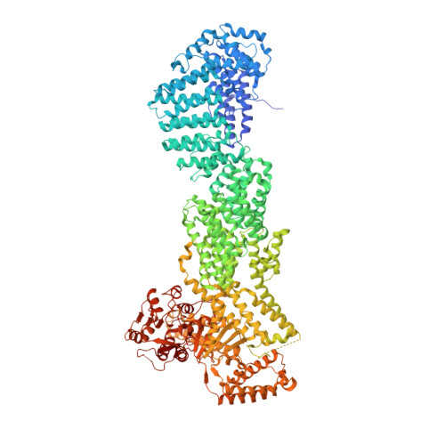
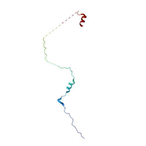
    Separase is a cysteine protease with a crucial role in the dissolution of cohesion among sister chromatids during chromosome segregation. In human tumours separase is overexpressed, making it a potential target for drug discovery. The protease activity of separase is strictly regulated by the inhibitor securin, which forms a tight complex with separase and may also stabilize this enzyme. Separases are large, 140-250-kilodalton enzymes, with an amino-terminal α-helical region and a carboxy-terminal caspase-like catalytic domain. Although crystal structures of the C-terminal two domains of separase and low-resolution electron microscopy reconstructions of the separase-securin complex have been reported, the atomic structures of full-length separase and especially the complex with securin are unknown. Here we report crystal structures at up to 2.6 Å resolution of the yeast Saccharomyces cerevisiae separase-securin complex. The α-helical region of separase (also known as Esp1) contains four domains (I-IV), and a substrate-binding domain immediately precedes the catalytic domain and has tight associations with it. The separase-securin complex assumes a highly elongated structure. Residues 258-373 of securin (Pds1), named the separase interaction segment, are primarily in an extended conformation and traverse the entire length of separase, interacting with all of its domains. Most importantly, residues 258-269 of securin are located in the separase active site, illuminating the mechanism of inhibition. Biochemical studies confirm the structural observations and indicate that contacts outside the separase active site are crucial for stabilizing the complex, thereby defining an important function for the helical region of separase.


  • Organizational Affiliation

    Department of Biological Sciences, Columbia University, New York, New York 10027, USA.


Macromolecules
Find similar proteins by:  (by identity cutoff)  |  3D Structure
Entity ID: 1
MoleculeChains Sequence LengthOrganismDetailsImage
Separin1,596Saccharomyces cerevisiae S288CMutation(s): 0 
Gene Names: ESP1YGR098C
EC: 3.4.22.49
UniProt
Find proteins for Q03018 (Saccharomyces cerevisiae (strain ATCC 204508 / S288c))
Explore Q03018 
Go to UniProtKB:  Q03018
Entity Groups  
Sequence Clusters30% Identity50% Identity70% Identity90% Identity95% Identity100% Identity
UniProt GroupQ03018
Sequence Annotations
Expand
  • Reference Sequence
Find similar proteins by:  (by identity cutoff)  |  3D Structure
Entity ID: 2
MoleculeChains Sequence LengthOrganismDetailsImage
Securin117Saccharomyces cerevisiae S288CMutation(s): 0 
Gene Names: PDS1YDR113CYD9727.08C
UniProt
Find proteins for P40316 (Saccharomyces cerevisiae (strain ATCC 204508 / S288c))
Explore P40316 
Go to UniProtKB:  P40316
Entity Groups  
Sequence Clusters30% Identity50% Identity70% Identity90% Identity95% Identity100% Identity
UniProt GroupP40316
Sequence Annotations
Expand
  • Reference Sequence
Experimental Data & Validation

Experimental Data

  • Method: X-RAY DIFFRACTION
  • Resolution: 2.60 Å
  • R-Value Free: 0.264 
  • R-Value Work: 0.221 
  • R-Value Observed: 0.222 
  • Space Group: P 32 2 1
Unit Cell:
Length ( Å )Angle ( ˚ )
a = 125.863α = 90
b = 125.863β = 90
c = 271.944γ = 120
Software Package:
Software NamePurpose
HKL-2000data scaling
PHENIXrefinement
PDB_EXTRACTdata extraction
HKL-2000data reduction
MR-Rosettaphasing
DENZOdata reduction
SCALEPACKdata scaling

Structure Validation

View Full Validation Report


Entry History & Funding Information

Deposition Data


Funding OrganizationLocationGrant Number
National Institutes of Health/National Institute of General Medical Sciences (NIH/NIGMS)United StatesR35GM118093
National Institutes of Health/Office of the DirectorUnited StatesS10OD012018

Revision History  (Full details and data files)

  • Version 1.0: 2017-02-08
    Type: Initial release
  • Version 1.1: 2017-02-15
    Changes: Database references
  • Version 1.2: 2017-02-22
    Changes: Database references
  • Version 1.3: 2017-09-27
    Changes: Author supporting evidence, Refinement description
  • Version 1.4: 2020-01-01
    Changes: Author supporting evidence
  • Version 1.5: 2023-10-04
    Changes: Data collection, Database references, Refinement description