5TUW

Crystal structure of Orange Carotenoid Protein with partial loss of 3'OH Echinenone chromophore


Experimental Data Snapshot

  • Method: X-RAY DIFFRACTION
  • Resolution: 2.30 Å
  • R-Value Free: 0.239 
  • R-Value Work: 0.185 
  • R-Value Observed: 0.186 

wwPDB Validation   3D Report Full Report


This is version 1.5 of the entry. See complete history


Literature

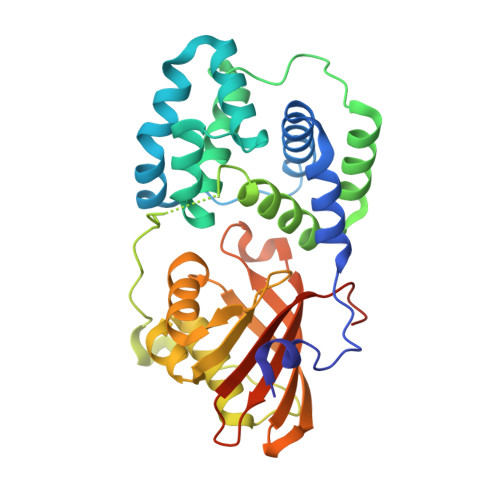
Photoactivation mechanism of a carotenoid-based photoreceptor.

Bandara, S.Ren, Z.Lu, L.Zeng, X.Shin, H.Zhao, K.H.Yang, X.

(2017) Proc Natl Acad Sci U S A 114: 6286-6291

  • DOI: https://doi.org/10.1073/pnas.1700956114
  • Primary Citation of Related Structures:  
    5TUW, 5TUX, 5TV0

  • PubMed Abstract: 

    Photoprotection is essential for efficient photosynthesis. Cyanobacteria have evolved a unique photoprotective mechanism mediated by a water-soluble carotenoid-based photoreceptor known as orange carotenoid protein (OCP). OCP undergoes large conformational changes in response to intense blue light, and the photoactivated OCP facilitates dissipation of excess energy via direct interaction with allophycocyanins at the phycobilisome core. However, the structural events leading up to the OCP photoactivation remain elusive at the molecular level. Here we present direct observations of light-induced structural changes in OCP captured by dynamic crystallography. Difference electron densities between the dark and illuminated states reveal widespread and concerted atomic motions that lead to altered protein-pigment interactions, displacement of secondary structures, and domain separation. Based on these crystallographic observations together with site-directed mutagenesis, we propose a molecular mechanism for OCP light perception, in which the photochemical property of a conjugated carbonyl group is exploited. We hypothesize that the OCP photoactivation starts with keto-enol tautomerization of the essential 4-keto group in the carotenoid, which disrupts the strong hydrogen bonds between the bent chromophore and the protein moiety. Subsequent structural changes trapped in the crystal lattice offer a high-resolution glimpse of the initial molecular events as OCP begins to transition from the orange-absorbing state to the active red-absorbing state.


  • Organizational Affiliation

    Department of Chemistry, University of Illinois at Chicago, Chicago, IL 60607.


Macromolecules
Find similar proteins by:  (by identity cutoff)  |  3D Structure
Entity ID: 1
MoleculeChains Sequence LengthOrganismDetailsImage
Orange carotenoid-binding protein
A, B, C, D, E
A, B, C, D, E, F
323Synechocystis sp. PCC 6803 substr. KazusaMutation(s): 0 
Gene Names: slr1963
UniProt
Find proteins for P74102 (Synechocystis sp. (strain PCC 6803 / Kazusa))
Explore P74102 
Go to UniProtKB:  P74102
Entity Groups  
Sequence Clusters30% Identity50% Identity70% Identity90% Identity95% Identity100% Identity
UniProt GroupP74102
Sequence Annotations
Expand
  • Reference Sequence
Small Molecules
Ligands 2 Unique
IDChains Name / Formula / InChI Key2D Diagram3D Interactions
EQ3
Query on EQ3

Download Ideal Coordinates CCD File 
H [auth A]
I [auth B]
L [auth C]
M [auth D]
O [auth E]
H [auth A],
I [auth B],
L [auth C],
M [auth D],
O [auth E],
P [auth F]
(3'R)-3'-hydroxy-beta,beta-caroten-4-one
C40 H54 O2
ZRCXVNZZDQGBQT-BANQPSJHSA-N
GOL
Query on GOL

Download Ideal Coordinates CCD File 
G [auth A],
J [auth C],
K [auth C],
N [auth E]
GLYCEROL
C3 H8 O3
PEDCQBHIVMGVHV-UHFFFAOYSA-N
Experimental Data & Validation

Experimental Data

  • Method: X-RAY DIFFRACTION
  • Resolution: 2.30 Å
  • R-Value Free: 0.239 
  • R-Value Work: 0.185 
  • R-Value Observed: 0.186 
  • Space Group: P 1
Unit Cell:
Length ( Å )Angle ( ˚ )
a = 82.947α = 89.79
b = 82.914β = 89.91
c = 87.438γ = 60.04
Software Package:
Software NamePurpose
PHENIXrefinement
HKL-2000data reduction
HKL-2000data scaling
PHENIXphasing

Structure Validation

View Full Validation Report


Entry History & Funding Information

Deposition Data


Funding OrganizationLocationGrant Number
National Institutes of Health/National Eye Institute (NIH/NEI)United StatesR01EY024363

Revision History  (Full details and data files)

  • Version 1.0: 2017-06-07
    Type: Initial release
  • Version 1.1: 2017-06-14
    Changes: Database references
  • Version 1.2: 2017-06-21
    Changes: Database references
  • Version 1.3: 2017-09-20
    Changes: Author supporting evidence
  • Version 1.4: 2019-12-11
    Changes: Author supporting evidence
  • Version 1.5: 2023-10-04
    Changes: Data collection, Database references, Refinement description, Structure summary