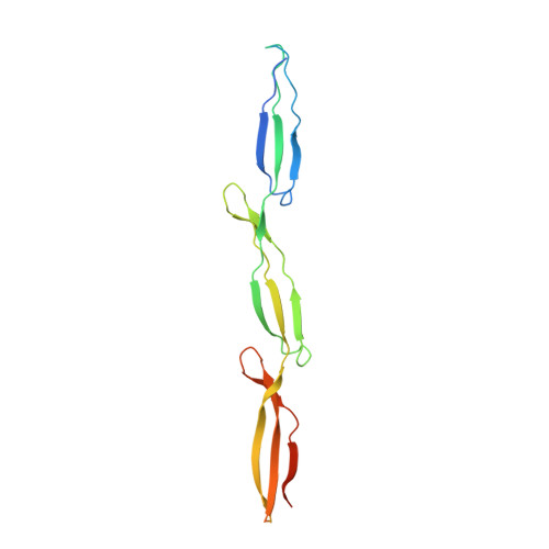
Functional consequences of B-repeat sequence variation in the staphylococcal biofilm protein Aap: deciphering the assembly code.
Shelton, C.L., Conrady, D.G., Herr, A.B.(2017) Biochem J 474: 427-443
- PubMed: 27872164
- DOI: https://doi.org/10.1042/BCJ20160675
- Primary Citation of Related Structures:
5TU7, 5TU8, 5TU9 - PubMed Abstract:
Staphylococcus epidermidis is an opportunistic pathogen that can form robust biofilms that render the bacteria resistant to antibiotic action and immune responses. Intercellular adhesion in S. epidermidis biofilms is mediated by the cell wall-associated accumulation-associated protein (Aap), via zinc-mediated self-assembly of its B-repeat region. This region contains up to 17 nearly identical sequence repeats, with each repeat assumed to be functionally equivalent. However, Aap B-repeats exist as two subtypes, defined by a cluster of consensus or variant amino acids. These variable residues are positioned near the zinc-binding (and dimerization) site and the stability determinant for the B-repeat fold. We have characterized four B-repeat constructs to assess the functional relevance of the two Aap B-repeat subtypes. Analytical ultracentrifugation experiments demonstrated that constructs with the variant sequence show reduced or absent Zn 2+ -induced dimerization. Likewise, circular dichroism thermal denaturation experiments showed that the variant sequence could significantly stabilize the fold, depending on its location within the construct. Crystal structures of three of the constructs revealed that the side chains from the variant sequence form an extensive bonding network that can stabilize the fold. Furthermore, altered distribution of charged residues between consensus and variant sequences changes the electrostatic potential in the vicinity of the Zn 2+ -binding site, providing a mechanistic explanation for the loss of zinc-induced dimerization in the variant constructs. These data suggest an assembly code that defines preferred oligomerization modes of the B-repeat region of Aap and a slip-grip model for initial contact followed by firm intercellular adhesion during biofilm formation.
Organizational Affiliation:
Program in Molecular Genetics, Biochemistry and Microbiology, University of Cincinnati College of Medicine, Cincinnati, OH 45267, U.S.A.


