


Crystal structure of Human Peroxisomal coenzyme A diphosphatase NUDT7
Srikannathasan, V.To be published.
Experimental Data Snapshot
wwPDB Validation 3D Report Full Report

Entity ID: 1 | |||||
|---|---|---|---|---|---|
| Molecule | Chains | Sequence Length | Organism | Details | Image |
| Peroxisomal coenzyme A diphosphatase NUDT7 | 236 | Homo sapiens | Mutation(s): 0 Gene Names: NUDT7 EC: 3.6.1 | 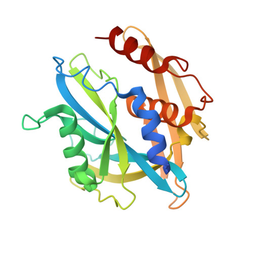 | |
UniProt & NIH Common Fund Data Resources | |||||
Find proteins for P0C024 (Homo sapiens) Explore P0C024 Go to UniProtKB: P0C024 | |||||
PHAROS: P0C024 GTEx: ENSG00000140876 | |||||
Entity Groups | |||||
| Sequence Clusters | 30% Identity50% Identity70% Identity90% Identity95% Identity100% Identity | ||||
| UniProt Group | P0C024 | ||||
Sequence AnnotationsExpand | |||||
| |||||
| Ligands 1 Unique | |||||
|---|---|---|---|---|---|
| ID | Chains | Name / Formula / InChI Key | 2D Diagram | 3D Interactions | |
| EDO Query on EDO | D [auth A] E [auth A] F [auth A] G [auth A] H [auth B] | 1,2-ETHANEDIOL C2 H6 O2 LYCAIKOWRPUZTN-UHFFFAOYSA-N |  | ||
| Length ( Å ) | Angle ( ˚ ) |
|---|---|
| a = 113.73 | α = 90 |
| b = 113.73 | β = 90 |
| c = 108.2 | γ = 120 |
| Software Name | Purpose |
|---|---|
| PHENIX | refinement |
| Aimless | data scaling |
| PDB_EXTRACT | data extraction |
| XDS | data reduction |
| MOLREP | phasing |
RCSB PDB (citation) is hosted by
RCSB PDB is a member of the

