5SWC

The structure of the beta-carbonic anhydrase CcaA


Experimental Data Snapshot

  • Method: X-RAY DIFFRACTION
  • Resolution: 1.45 Å
  • R-Value Free: 0.146 
  • R-Value Work: 0.131 
  • R-Value Observed: 0.131 

wwPDB Validation   3D Report Full Report


This is version 1.4 of the entry. See complete history


Literature

The structure, kinetics and interactions of the beta-carboxysomal beta-carbonic anhydrase, CcaA.

McGurn, L.D.Moazami-Goudarzi, M.White, S.A.Suwal, T.Brar, B.Tang, J.Q.Espie, G.S.Kimber, M.S.

(2016) Biochem J 473: 4559-4572

  • DOI: https://doi.org/10.1042/BCJ20160773
  • Primary Citation of Related Structures:  
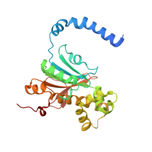
    5SWC

  • PubMed Abstract: 

    CcaA is a β-carbonic anhydrase (CA) that is a component of the carboxysomes of a subset of β-cyanobacteria. This protein, which has a characteristic C-terminal extension of unknown function, is recruited to the carboxysome via interactions with CcmM, which is itself a γ-CA homolog with enzymatic activity in many, but not all cyanobacteria. We have determined the structure of CcaA from Synechocystis sp. PCC 6803 at 1.45 Å. In contrast with the dimer-of-dimers organization of most bacterial β-CAs, or the loose dimer-of-dimers-of-dimers organization found in the plant enzymes, CcaA shows a well-packed trimer-of-dimers organization. The proximal part of the characteristic C-terminal extension is ordered by binding at a site that passes through the two-fold symmetry axis shared with an adjacent dimer; as a result, only one of a pair of converging termini can be ordered at any given time. Docking in Rosetta failed to find well-packed solutions, indicating that formation of the CcaA/CcmM complex probably requires significant backbone movements in at least one of the binding partners. Surface plasmon resonance experiments showed that CcaA forms a complex with CcmM with sub-picomolar affinity, with contributions from residues in CcmM's αA helix and CcaA's C-terminal tail. Catalytic characterization showed CcaA to be among the least active β-CAs characterized to date, with activity comparable with the γ-CA, CcmM, it either complements or replaces. Intriguingly, the C-terminal tail appears to partly inhibit activity, possibly indicating a role in minimizing the activity of unencapsulated enzyme.


  • Organizational Affiliation

    Department of Molecular and Cellular Biology, University of Guelph, Guelph, Ontario, Canada N1G2W1.


Macromolecules
Find similar proteins by:  (by identity cutoff)  |  3D Structure
Entity ID: 1
MoleculeChains Sequence LengthOrganismDetailsImage
Carbonic anhydrase
A, B, C, D, E
A, B, C, D, E, F
239Synechocystis sp. PCC 6803 substr. KazusaMutation(s): 0 
Gene Names: icfAslr1347
EC: 4.2.1.1
UniProt
Find proteins for Q54735 (Synechocystis sp. (strain PCC 6803 / Kazusa))
Explore Q54735 
Go to UniProtKB:  Q54735
Entity Groups  
Sequence Clusters30% Identity50% Identity70% Identity90% Identity95% Identity100% Identity
UniProt GroupQ54735
Sequence Annotations
Expand
  • Reference Sequence
Small Molecules
Ligands 3 Unique
IDChains Name / Formula / InChI Key2D Diagram3D Interactions
ZN
Query on ZN

Download Ideal Coordinates CCD File 
BA [auth E]
G [auth A]
JA [auth F]
M [auth B]
S [auth C]
BA [auth E],
G [auth A],
JA [auth F],
M [auth B],
S [auth C],
Y [auth D]
ZINC ION
Zn
PTFCDOFLOPIGGS-UHFFFAOYSA-N
FMT
Query on FMT

Download Ideal Coordinates CCD File 
AA [auth D]
EA [auth E]
FA [auth E]
GA [auth E]
HA [auth E]
AA [auth D],
EA [auth E],
FA [auth E],
GA [auth E],
HA [auth E],
IA [auth E],
J [auth A],
K [auth A],
KA [auth F],
L [auth A],
LA [auth F],
N [auth B],
O [auth B],
P [auth B],
Q [auth B],
R [auth B],
U [auth C],
V [auth C],
W [auth C],
X [auth C],
Z [auth D]
FORMIC ACID
C H2 O2
BDAGIHXWWSANSR-UHFFFAOYSA-N
CL
Query on CL

Download Ideal Coordinates CCD File 
CA [auth E],
DA [auth E],
H [auth A],
I [auth A],
T [auth C]
CHLORIDE ION
Cl
VEXZGXHMUGYJMC-UHFFFAOYSA-M
Experimental Data & Validation

Experimental Data

  • Method: X-RAY DIFFRACTION
  • Resolution: 1.45 Å
  • R-Value Free: 0.146 
  • R-Value Work: 0.131 
  • R-Value Observed: 0.131 
  • Space Group: I 4
Unit Cell:
Length ( Å )Angle ( ˚ )
a = 202.95α = 90
b = 202.95β = 90
c = 70.51γ = 90
Software Package:
Software NamePurpose
PHENIXrefinement
XDSdata reduction
XSCALEdata scaling
PHASERphasing

Structure Validation

View Full Validation Report


Entry History & Funding Information

Deposition Data


Funding OrganizationLocationGrant Number
Natural Sciences and Engineering Research Council (NSERC, Canada)Canada04045-2015

Revision History  (Full details and data files)

  • Version 1.0: 2016-10-26
    Type: Initial release
  • Version 1.1: 2016-12-28
    Changes: Database references
  • Version 1.2: 2017-09-27
    Changes: Author supporting evidence
  • Version 1.3: 2020-01-08
    Changes: Author supporting evidence
  • Version 1.4: 2023-10-04
    Changes: Data collection, Database references, Derived calculations, Refinement description