Structural Transitions of the Conserved and Metastable Hantaviral Glycoprotein Envelope.
Rissanen, I., Stass, R., Zeltina, A., Li, S., Hepojoki, J., Harlos, K., Gilbert, R.J.C., Huiskonen, J.T., Bowden, T.A.(2017) J Virol 91
- PubMed: 28835498
- DOI: https://doi.org/10.1128/JVI.00378-17
- Primary Citation of Related Structures:
5OPG - PubMed Abstract:
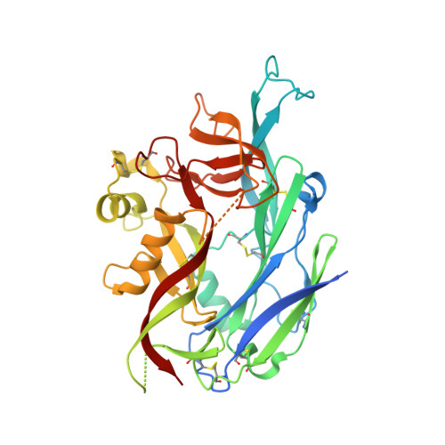
Hantaviruses are zoonotic pathogens that cause severe hemorrhagic fever and pulmonary syndrome. The outer membrane of the hantavirus envelope displays a lattice of two glycoproteins, Gn and Gc, which orchestrate host cell recognition and entry. Here, we describe the crystal structure of the Gn glycoprotein ectodomain from the Asiatic Hantaan virus (HTNV), the most prevalent pathogenic hantavirus. Structural overlay analysis reveals that the HTNV Gn fold is highly similar to the Gn of Puumala virus (PUUV), a genetically and geographically distinct and less pathogenic hantavirus found predominantly in northeastern Europe, confirming that the hantaviral Gn fold is architecturally conserved across hantavirus clades. Interestingly, HTNV Gn crystallized at acidic pH, in a compact tetrameric configuration distinct from the organization at neutral pH. Analysis of the Gn, both in solution and in the context of the virion, confirms the pH-sensitive oligomeric nature of the glycoprotein, indicating that the hantaviral Gn undergoes structural transitions during host cell entry. These data allow us to present a structural model for how acidification during endocytic uptake of the virus triggers the dissociation of the metastable Gn-Gc lattice to enable insertion of the Gc-resident hydrophobic fusion loops into the host cell membrane. Together, these data reveal the dynamic plasticity of the structurally conserved hantaviral surface. IMPORTANCE Although outbreaks of Korean hemorrhagic fever were first recognized during the Korean War (1950 to 1953), it was not until 1978 that they were found to be caused by Hantaan virus (HTNV), the most prevalent pathogenic hantavirus. Here, we describe the crystal structure of HTNV envelope glycoprotein Gn, an integral component of the Gn-Gc glycoprotein spike complex responsible for host cell entry. HTNV Gn is structurally conserved with the Gn of a genetically and geographically distal hantavirus, Puumala virus, indicating that the observed α/β fold is well preserved across the Hantaviridae family. The combination of our crystal structure with solution state analysis of recombinant protein and electron cryo-microscopy of acidified hantavirus allows us to propose a model for endosome-induced reorganization of the hantaviral glycoprotein lattice. This provides a molecular-level rationale for the exposure of the hydrophobic fusion loops on the Gc, a process required for fusion of viral and cellular membranes.
Organizational Affiliation:
Division of Structural Biology, Wellcome Trust Centre for Human Genetics, University of Oxford, Oxford, United Kingdom.