5O44

Crystal structure of unbranched mixed tri-Ubiquitin chain containing K48 and K63 linkages.


Experimental Data Snapshot

  • Method: X-RAY DIFFRACTION
  • Resolution: 3.14 Å
  • R-Value Free: 0.254 
  • R-Value Work: 0.217 
  • R-Value Observed: 0.219 

wwPDB Validation   3D Report Full Report


This is version 1.4 of the entry. See complete history


Literature

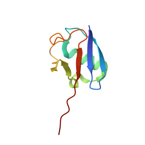
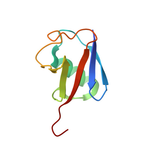
The Crystal Structure and Conformations of an Unbranched Mixed Tri-Ubiquitin Chain Containing K48 and K63 Linkages.

Padala, P.Soudah, N.Giladi, M.Haitin, Y.Isupov, M.N.Wiener, R.

(2017) J Mol Biol 429: 3801-3813

  • DOI: https://doi.org/10.1016/j.jmb.2017.10.027
  • Primary Citation of Related Structures:  
    5O44

  • PubMed Abstract: 

    The ability of ubiquitin to function in a wide range of cellular processes is ascribed to its capacity to cause a diverse spectrum of modifications. While a target protein can be modified with monoubiquitin, it can also be modified with ubiquitin chains. The latter include seven types of homotypic chains as well as mixed ubiquitin chains. In a mixed chain, not all the isopeptide bonds are restricted to a specific lysine of ubiquitin, resulting in a chain possessing more than one type of linkage. While structural characterization of homotypic chains has been well elucidated, less is known about mixed chains. Here we present the crystal structure of a mixed tri-ubiquitin chain at 3.1-Å resolution. In the structure, the proximal ubiquitin is connected to the middle ubiquitin via K48 and these two ubiquitins adopt a compact structure as observed in K48 di-ubiquitin. The middle ubiquitin links to the distal ubiquitin via its K63 and these ubiquitins adopt two conformations, suggesting a flexible structure. Using small-angle X-ray scattering, we unexpectedly found differences between the conformational ensembles of the above tri-ubiquitin chains and chains possessing the same linkages but in the reverse order. In addition, cleavage of the K48 linkage by DUB is faster if this linkage is at the distal end. Taken together, our results suggest that in mixed chains, not only the type of the linkages but also their sequence determine the structural and functional properties of the chain.


  • Organizational Affiliation

    Department of Biochemistry and Molecular Biology, the Institute for Medical Research Israel-Canada, Hebrew University-Hadassah Medical School, Jerusalem 91120, Israel.


Macromolecules
Find similar proteins by:  (by identity cutoff)  |  3D Structure
Entity ID: 1
MoleculeChains Sequence LengthOrganismDetailsImage
Ubiquitin
A, E
76Musca domesticaMutation(s): 1 
UniProt
Find proteins for Q45TR8 (Musca domestica)
Explore Q45TR8 
Go to UniProtKB:  Q45TR8
Entity Groups  
Sequence Clusters30% Identity50% Identity70% Identity90% Identity95% Identity100% Identity
UniProt GroupQ45TR8
Sequence Annotations
Expand
  • Reference Sequence
Find similar proteins by:  (by identity cutoff)  |  3D Structure
Entity ID: 2
MoleculeChains Sequence LengthOrganismDetailsImage
Polyubiquitin-BB [auth C],
D [auth B]
74Homo sapiensMutation(s): 0 
Gene Names: UBB
UniProt & NIH Common Fund Data Resources
Find proteins for P0CG47 (Homo sapiens)
Explore P0CG47 
Go to UniProtKB:  P0CG47
PHAROS:  P0CG47
GTEx:  ENSG00000170315 
Entity Groups  
Sequence Clusters30% Identity50% Identity70% Identity90% Identity95% Identity100% Identity
UniProt GroupP0CG47
Sequence Annotations
Expand
  • Reference Sequence
Find similar proteins by:  (by identity cutoff)  |  3D Structure
Entity ID: 3
MoleculeChains Sequence LengthOrganismDetailsImage
Polyubiquitin-BC [auth D],
F
76Homo sapiensMutation(s): 1 
Gene Names: UBB
UniProt & NIH Common Fund Data Resources
Find proteins for P0CG47 (Homo sapiens)
Explore P0CG47 
Go to UniProtKB:  P0CG47
PHAROS:  P0CG47
GTEx:  ENSG00000170315 
Entity Groups  
Sequence Clusters30% Identity50% Identity70% Identity90% Identity95% Identity100% Identity
UniProt GroupP0CG47
Sequence Annotations
Expand
  • Reference Sequence
Small Molecules
Ligands 2 Unique
IDChains Name / Formula / InChI Key2D Diagram3D Interactions
SO4
Query on SO4

Download Ideal Coordinates CCD File 
G [auth A]
H [auth A]
J [auth C]
K [auth C]
M [auth B]
G [auth A],
H [auth A],
J [auth C],
K [auth C],
M [auth B],
N [auth B],
O [auth E],
P [auth E],
Q [auth E]
SULFATE ION
O4 S
QAOWNCQODCNURD-UHFFFAOYSA-L
MG
Query on MG

Download Ideal Coordinates CCD File 
I [auth A],
L [auth D],
R [auth E]
MAGNESIUM ION
Mg
JLVVSXFLKOJNIY-UHFFFAOYSA-N
Experimental Data & Validation

Experimental Data

  • Method: X-RAY DIFFRACTION
  • Resolution: 3.14 Å
  • R-Value Free: 0.254 
  • R-Value Work: 0.217 
  • R-Value Observed: 0.219 
  • Space Group: P 61 2 2
Unit Cell:
Length ( Å )Angle ( ˚ )
a = 110.767α = 90
b = 110.767β = 90
c = 417.004γ = 120
Software Package:
Software NamePurpose
REFMACrefinement
xia2data reduction
Aimlessdata scaling
MOLREPphasing
DMphasing
XDSdata reduction

Structure Validation

View Full Validation Report


Entry History & Funding Information

Deposition Data


Funding OrganizationLocationGrant Number
Israel--

Revision History  (Full details and data files)

  • Version 1.0: 2017-11-08
    Type: Initial release
  • Version 1.1: 2017-11-15
    Changes: Database references
  • Version 1.2: 2017-12-06
    Changes: Database references
  • Version 1.3: 2019-05-08
    Changes: Advisory, Data collection, Derived calculations
  • Version 1.4: 2024-01-17
    Changes: Data collection, Database references, Refinement description