5MV5

Structure of deformed wing virus, a honeybee pathogen


Experimental Data Snapshot

  • Method: ELECTRON MICROSCOPY
  • Resolution: 3.10 Å
  • Aggregation State: PARTICLE 
  • Reconstruction Method: SINGLE PARTICLE 

wwPDB Validation   3D Report Full Report


This is version 1.2 of the entry. See complete history


Literature

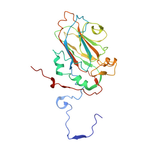
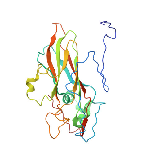
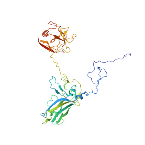
Structure of deformed wing virus, a major honey bee pathogen.

Skubnik, K.Novacek, J.Fuzik, T.Pridal, A.Paxton, R.J.Plevka, P.

(2017) Proc Natl Acad Sci U S A 114: 3210-3215

  • DOI: https://doi.org/10.1073/pnas.1615695114
  • Primary Citation of Related Structures:  
    5G51, 5G52, 5L7Q, 5L8Q, 5MUP, 5MV5, 5MV6

  • PubMed Abstract: 

    The worldwide population of western honey bees ( Apis mellifera ) is under pressure from habitat loss, environmental stress, and pathogens, particularly viruses that cause lethal epidemics. Deformed wing virus (DWV) from the family Iflaviridae , together with its vector, the mite Varroa destructor , is likely the major threat to the world's honey bees. However, lack of knowledge of the atomic structures of iflaviruses has hindered the development of effective treatments against them. Here, we present the virion structures of DWV determined to a resolution of 3.1 Å using cryo-electron microscopy and 3.8 Å by X-ray crystallography. The C-terminal extension of capsid protein VP3 folds into a globular protruding (P) domain, exposed on the virion surface. The P domain contains an Asp-His-Ser catalytic triad that is, together with five residues that are spatially close, conserved among iflaviruses. These residues may participate in receptor binding or provide the protease, lipase, or esterase activity required for entry of the virus into a host cell. Furthermore, nucleotides of the DWV RNA genome interact with VP3 subunits. The capsid protein residues involved in the RNA binding are conserved among honey bee iflaviruses, suggesting a putative role of the genome in stabilizing the virion or facilitating capsid assembly. Identifying the RNA-binding and putative catalytic sites within the DWV virion structure enables future analyses of how DWV and other iflaviruses infect insect cells and also opens up possibilities for the development of antiviral treatments.


  • Organizational Affiliation

    Structural Virology, Central European Institute of Technology, Masaryk University, 62500 Brno, Czech Republic.


Macromolecules
Find similar proteins by:  (by identity cutoff)  |  3D Structure
Entity ID: 1
MoleculeChains Sequence LengthOrganismDetailsImage
VP1258Deformed wing virusMutation(s): 0 
UniProt
Find proteins for L0CTV4 (Deformed wing virus)
Explore L0CTV4 
Go to UniProtKB:  L0CTV4
Entity Groups  
Sequence Clusters30% Identity50% Identity70% Identity90% Identity95% Identity100% Identity
UniProt GroupL0CTV4
Sequence Annotations
Expand
  • Reference Sequence
Find similar proteins by:  (by identity cutoff)  |  3D Structure
Entity ID: 2
MoleculeChains Sequence LengthOrganismDetailsImage
VP2253Deformed wing virusMutation(s): 0 
UniProt
Find proteins for Q7TG18 (Deformed wing virus)
Explore Q7TG18 
Go to UniProtKB:  Q7TG18
Entity Groups  
Sequence Clusters30% Identity50% Identity70% Identity90% Identity95% Identity100% Identity
UniProt GroupQ7TG18
Sequence Annotations
Expand
  • Reference Sequence
Find similar proteins by:  (by identity cutoff)  |  3D Structure
Entity ID: 3
MoleculeChains Sequence LengthOrganismDetailsImage
VP3416Deformed wing virusMutation(s): 0 
UniProt
Find proteins for Q7TG18 (Deformed wing virus)
Explore Q7TG18 
Go to UniProtKB:  Q7TG18
Entity Groups  
Sequence Clusters30% Identity50% Identity70% Identity90% Identity95% Identity100% Identity
UniProt GroupQ7TG18
Sequence Annotations
Expand
  • Reference Sequence
Small Molecules
Ligands 1 Unique
IDChains Name / Formula / InChI Key2D Diagram3D Interactions
U
Query on U

Download Ideal Coordinates CCD File 
D [auth C]URIDINE-5'-MONOPHOSPHATE
C9 H13 N2 O9 P
DJJCXFVJDGTHFX-XVFCMESISA-N
Experimental Data & Validation

Experimental Data

  • Method: ELECTRON MICROSCOPY
  • Resolution: 3.10 Å
  • Aggregation State: PARTICLE 
  • Reconstruction Method: SINGLE PARTICLE 
EM Software:
TaskSoftware PackageVersion
MODEL REFINEMENTPHENIX
RECONSTRUCTIONRELION1.4

Structure Validation

View Full Validation Report


Entry History & Funding Information

Deposition Data


Funding OrganizationLocationGrant Number
European Research Council355855
European Molecular Biology Organization3041

Revision History  (Full details and data files)

  • Version 1.0: 2017-04-05
    Type: Initial release
  • Version 1.1: 2017-08-02
    Changes: Data collection, Refinement description
  • Version 1.2: 2017-08-30
    Changes: Data collection