5MU1

UDP-Glucose Glycoprotein Glucosyltransferase from Chaetomium thermophilum soaked with K2PtI6


Experimental Data Snapshot

  • Method: X-RAY DIFFRACTION
  • Resolution: 3.48 Å
  • R-Value Free: 0.252 
  • R-Value Work: 0.237 
  • R-Value Observed: 0.238 

wwPDB Validation   3D Report Full Report


This is version 2.1 of the entry. See complete history


Literature

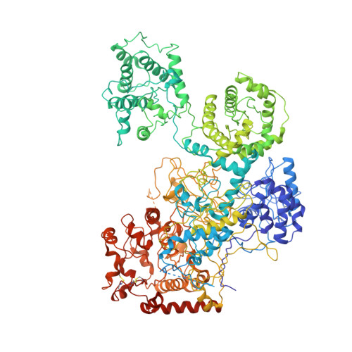
Interdomain conformational flexibility underpins the activity of UGGT, the eukaryotic glycoprotein secretion checkpoint.

Roversi, P.Marti, L.Caputo, A.T.Alonzi, D.S.Hill, J.C.Dent, K.C.Kumar, A.Levasseur, M.D.Lia, A.Waksman, T.Basu, S.Soto Albrecht, Y.Qian, K.McIvor, J.P.Lipp, C.B.Siliqi, D.Vasiljevic, S.Mohammed, S.Lukacik, P.Walsh, M.A.Santino, A.Zitzmann, N.

(2017) Proc Natl Acad Sci U S A 114: 8544-8549

  • DOI: https://doi.org/10.1073/pnas.1703682114
  • Primary Citation of Related Structures:  
    5MU1, 5MZO, 5N2J, 5NV4

  • PubMed Abstract: 

    Glycoproteins traversing the eukaryotic secretory pathway begin life in the endoplasmic reticulum (ER), where their folding is surveyed by the 170-kDa UDP-glucose:glycoprotein glucosyltransferase (UGGT). The enzyme acts as the single glycoprotein folding quality control checkpoint: it selectively reglucosylates misfolded glycoproteins, promotes their association with ER lectins and associated chaperones, and prevents premature secretion from the ER. UGGT has long resisted structural determination and sequence-based domain boundary prediction. Questions remain on how this single enzyme can flag misfolded glycoproteins of different sizes and shapes for ER retention and how it can span variable distances between the site of misfold and a glucose-accepting N-linked glycan on the same glycoprotein. Here, crystal structures of a full-length eukaryotic UGGT reveal four thioredoxin-like (TRXL) domains arranged in a long arc that terminates in two β-sandwiches tightly clasping the glucosyltransferase domain. The fold of the molecule is topologically complex, with the first β-sandwich and the fourth TRXL domain being encoded by nonconsecutive stretches of sequence. In addition to the crystal structures, a 15-Å cryo-EM reconstruction reveals interdomain flexibility of the TRXL domains. Double cysteine point mutants that engineer extra interdomain disulfide bridges rigidify the UGGT structure and exhibit impaired activity. The intrinsic flexibility of the TRXL domains of UGGT may therefore endow the enzyme with the promiscuity needed to recognize and reglucosylate its many different substrates and/or enable reglucosylation of N-linked glycans situated at variable distances from the site of misfold.


  • Organizational Affiliation

    Oxford Glycobiology Institute, Department of Biochemistry, University of Oxford, Oxford OX1 3QU, United Kingdom; pietro.roversi@bioch.ox.ac.uk nicole.zitzmann@bioch.ox.ac.uk.


Macromolecules
Find similar proteins by:  (by identity cutoff)  |  3D Structure
Entity ID: 1
MoleculeChains Sequence LengthOrganismDetailsImage
UDP-glucose-glycoprotein glucosyltransferase-like protein1,494Thermochaetoides thermophila DSM 1495Mutation(s): 0 
Gene Names: CTHT_0048990
UniProt
Find proteins for G0SB58 (Chaetomium thermophilum (strain DSM 1495 / CBS 144.50 / IMI 039719))
Explore G0SB58 
Go to UniProtKB:  G0SB58
Entity Groups  
Sequence Clusters30% Identity50% Identity70% Identity90% Identity95% Identity100% Identity
UniProt GroupG0SB58
Sequence Annotations
Expand
  • Reference Sequence
Oligosaccharides

Help

Entity ID: 2
MoleculeChains Length2D Diagram Glycosylation3D Interactions
alpha-D-mannopyranose-(1-3)-alpha-D-mannopyranose-(1-3)-[alpha-D-mannopyranose-(1-6)-alpha-D-mannopyranose-(1-6)]beta-D-mannopyranose-(1-4)-2-acetamido-2-deoxy-beta-D-glucopyranose-(1-4)-2-acetamido-2-deoxy-beta-D-glucopyranose
B
7N-Glycosylation
Glycosylation Resources
GlyTouCan:  G84881ST
GlyCosmos:  G84881ST
GlyGen:  G84881ST
Entity ID: 3
MoleculeChains Length2D Diagram Glycosylation3D Interactions
alpha-D-mannopyranose-(1-3)-[alpha-D-mannopyranose-(1-6)]beta-D-mannopyranose-(1-4)-2-acetamido-2-deoxy-beta-D-glucopyranose-(1-4)-2-acetamido-2-deoxy-beta-D-glucopyranose
C
5N-Glycosylation
Glycosylation Resources
GlyTouCan:  G22768VO
GlyCosmos:  G22768VO
GlyGen:  G22768VO
Entity ID: 4
MoleculeChains Length2D Diagram Glycosylation3D Interactions
alpha-D-mannopyranose-(1-6)-alpha-D-mannopyranose-(1-3)-beta-D-mannopyranose-(1-4)-2-acetamido-2-deoxy-beta-D-glucopyranose-(1-4)-2-acetamido-2-deoxy-beta-D-glucopyranose
D
5N-Glycosylation
Glycosylation Resources
GlyTouCan:  G77367MI
GlyCosmos:  G77367MI
GlyGen:  G77367MI
Entity ID: 5
MoleculeChains Length2D Diagram Glycosylation3D Interactions
alpha-D-mannopyranose-(1-3)-[alpha-D-mannopyranose-(1-6)]alpha-D-mannopyranose-(1-6)-alpha-D-mannopyranose-(1-3)-beta-D-mannopyranose-(1-4)-2-acetamido-2-deoxy-beta-D-glucopyranose-(1-4)-2-acetamido-2-deoxy-beta-D-glucopyranose
E
7N-Glycosylation
Glycosylation Resources
GlyTouCan:  G11568OK
GlyCosmos:  G11568OK
GlyGen:  G11568OK
Small Molecules
Ligands 3 Unique
IDChains Name / Formula / InChI Key2D Diagram3D Interactions
PT
Query on PT

Download Ideal Coordinates CCD File 
I [auth A]
J [auth A]
O [auth A]
R [auth A]
V [auth A]
I [auth A],
J [auth A],
O [auth A],
R [auth A],
V [auth A],
Y [auth A]
PLATINUM (II) ION
Pt
HRGDZIGMBDGFTC-UHFFFAOYSA-N
IOD
Query on IOD

Download Ideal Coordinates CCD File 
F [auth A]
G [auth A]
H [auth A]
K [auth A]
L [auth A]
F [auth A],
G [auth A],
H [auth A],
K [auth A],
L [auth A],
M [auth A],
N [auth A],
P [auth A],
Q [auth A],
S [auth A],
T [auth A],
U [auth A],
W [auth A],
X [auth A]
IODIDE ION
I
XMBWDFGMSWQBCA-UHFFFAOYSA-M
CA
Query on CA

Download Ideal Coordinates CCD File 
Z [auth A]CALCIUM ION
Ca
BHPQYMZQTOCNFJ-UHFFFAOYSA-N
Experimental Data & Validation

Experimental Data

  • Method: X-RAY DIFFRACTION
  • Resolution: 3.48 Å
  • R-Value Free: 0.252 
  • R-Value Work: 0.237 
  • R-Value Observed: 0.238 
  • Space Group: P 61
Unit Cell:
Length ( Å )Angle ( ˚ )
a = 116.95α = 90
b = 116.95β = 90
c = 301.491γ = 120
Software Package:
Software NamePurpose
BUSTERrefinement
XDSdata reduction
Aimlessdata scaling
autoSHARPphasing

Structure Validation

View Full Validation Report


Entry History & Funding Information

Deposition Data


Funding OrganizationLocationGrant Number
Wellcome TrustUnited Kingdom097300/Z/11/Z
Wellcome TrustUnited Kingdom106272/Z/14/Z

Revision History  (Full details and data files)

  • Version 1.0: 2017-07-26
    Type: Initial release
  • Version 1.1: 2017-08-02
    Changes: Database references
  • Version 1.2: 2017-08-16
    Changes: Database references
  • Version 1.3: 2020-04-08
    Changes: Data collection
  • Version 1.4: 2020-06-03
    Changes: Derived calculations
  • Version 2.0: 2020-07-29
    Type: Remediation
    Reason: Carbohydrate remediation
    Changes: Atomic model, Data collection, Derived calculations, Structure summary
  • Version 2.1: 2023-03-08
    Changes: Database references, Derived calculations, Structure summary