Nigritoxin is a bacterial toxin for crustaceans and insects.
Labreuche, Y., Chenivesse, S., Jeudy, A., Le Panse, S., Boulo, V., Ansquer, D., Pages, S., Givaudan, A., Czjzek, M., Le Roux, F.(2017) Nat Commun 8: 1248-1248
- PubMed: 29093459
- DOI: https://doi.org/10.1038/s41467-017-01445-z
- Primary Citation of Related Structures:
5M41 - PubMed Abstract:
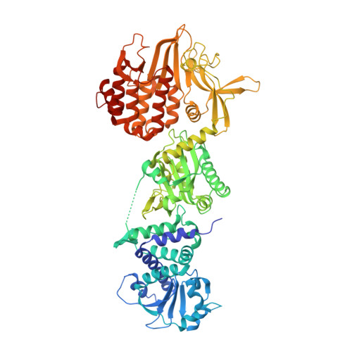
The Tetraconata (Pancrustacea) concept proposes that insects are more closely related to aquatic crustaceans than to terrestrial centipedes or millipedes. The question therefore arises whether insects have kept crustacean-specific genetic traits that could be targeted by specific toxins. Here we show that a toxin (nigritoxin), originally identified in a bacterial pathogen of shrimp, is lethal for organisms within the Tetraconata and non-toxic to other animals. X-ray crystallography reveals that nigritoxin possesses a new protein fold of the α/β type. The nigritoxin N-terminal domain is essential for cellular translocation and likely encodes specificity for Tetraconata. Once internalized by eukaryotic cells, nigritoxin induces apoptotic cell death through structural features that are localized in the C-terminal domain of the protein. We propose that nigritoxin will be an effective means to identify a Tetraconata evolutionarily conserved pathway and speculate that nigritoxin holds promise as an insecticidal protein.
Organizational Affiliation:
Ifremer, Unité Physiologie Fonctionnelle des Organismes Marins, ZI de la Pointe du Diable, CS 10070, F-29280, Plouzané, France.