5M2F

Crystal structure of human AKR1B10 complexed with NADP+ and the synthetic retinoid UVI2008


Experimental Data Snapshot

  • Method: X-RAY DIFFRACTION
  • Resolution: 1.50 Å
  • R-Value Free: 0.241 
  • R-Value Work: 0.210 
  • R-Value Observed: 0.212 

wwPDB Validation   3D Report Full Report


Ligand Structure Quality Assessment 


This is version 1.2 of the entry. See complete history


Literature

Structural basis for the inhibition of AKR1B10 by the C3 brominated TTNPB derivative UVI2008.

Ruiz, F.X.Crespo, I.Alvarez, S.Porte, S.Gimenez-Dejoz, J.Cousido-Siah, A.Mitschler, A.de Lera, A.R.Pares, X.Podjarny, A.Farres, J.

(2017) Chem Biol Interact 276: 174-181

  • DOI: https://doi.org/10.1016/j.cbi.2017.01.026
  • Primary Citation of Related Structures:  
    5M2F

  • PubMed Abstract: 

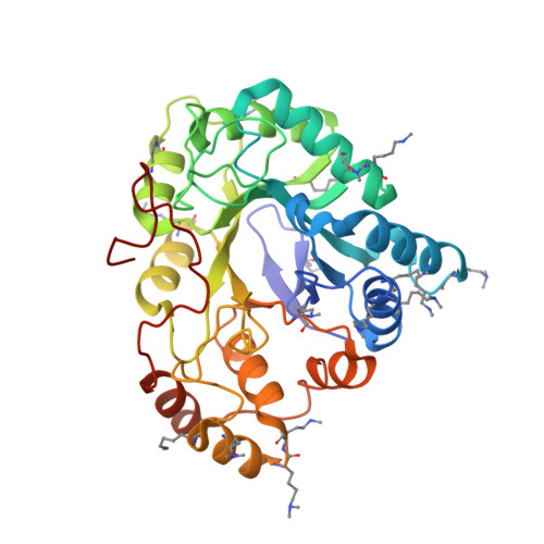
    UVI2008, a retinoic acid receptor (RAR) β/γ agonist originated from C3 bromine addition to the parent RAR pan-agonist 4-[(E)-2-(5,6,7,8-tetrahydro-5,5,8,8-tetramethyl-2-naphthalenyl)-1-propenyl]benzoic acid (TTNPB), is also a selective inhibitor of aldo-keto reductase family member 1B10 (AKR1B10). Thus, it might become a lead drug for the design of compounds targeting both activities, as an AKR1B10 inhibitor and RAR agonist, which could constitute a novel therapeutic approach against cancer and skin-related diseases. Herein, the X-ray structure of the methylated Lys125Arg/Val301Leu AKR1B10 (i.e. AKME2MU) holoenzyme in complex with UVI2008 was determined at 1.5 Å resolution, providing an explanation for UVI2008 selectivity against AKR1B10 (IC 50  = 6.1 μM) over the closely related aldose reductase (AR, IC 50  = 70 μM). The carboxylic acid group of UVI2008 is located in the anion-binding pocket, at hydrogen-bond distance of catalytically important residues Tyr49 and His111. The inhibitor bromine atom can only fit in the wider active site of AKR1B10, mainly because of the native Trp112 side-chain orientation, not possible in AR. In AKR1B10, Trp112 native conformation, and thus UVI2008 binding, is facilitated through interaction with Gln114. IC 50 analysis of the corresponding Thr113Gln mutant in AR confirmed this hypothesis. The elucidation of the binding mode of UVI2008 to AKR1B10, along with the previous studies on the retinoid specificity of AKR1B10 and on the stilbene retinoid scaffold conforming UVI2008, could indeed be used to foster the drug design of bifunctional antiproliferative compounds.


  • Organizational Affiliation

    Institut de Génétique et de Biologie Moléculaire et Cellulaire, 1 rue Laurent Fries, 67404 Illkirch Cedex, France. Electronic address: fxavier.ruiz@gmail.com.


Macromolecules
Find similar proteins by:  (by identity cutoff)  |  3D Structure
Entity ID: 1
MoleculeChains Sequence LengthOrganismDetailsImage
Aldo-keto reductase family 1 member B10A [auth X]316Homo sapiensMutation(s): 2 
Gene Names: AKR1B10AKR1B11
EC: 1.1.1
UniProt & NIH Common Fund Data Resources
Find proteins for O60218 (Homo sapiens)
Explore O60218 
Go to UniProtKB:  O60218
PHAROS:  O60218
GTEx:  ENSG00000198074 
Entity Groups  
Sequence Clusters30% Identity50% Identity70% Identity90% Identity95% Identity100% Identity
UniProt GroupO60218
Sequence Annotations
Expand
  • Reference Sequence
Small Molecules
Ligands 3 Unique
IDChains Name / Formula / InChI Key2D Diagram3D Interactions
NAP
Query on NAP

Download Ideal Coordinates CCD File 
B [auth X]NADP NICOTINAMIDE-ADENINE-DINUCLEOTIDE PHOSPHATE
C21 H28 N7 O17 P3
XJLXINKUBYWONI-NNYOXOHSSA-N
UV8
Query on UV8

Download Ideal Coordinates CCD File 
C [auth X]3-bromo-4-[(1E)-2-(5,5,8,8-tetramethyl-5,6,7,8-tetrahydronaphthalen-2-yl)prop-1-en-1-yl]benzoic acid
C24 H27 Br O2
CTUBWRWXDXMLOB-UHFFFAOYSA-N
EDO
Query on EDO

Download Ideal Coordinates CCD File 
D [auth X]1,2-ETHANEDIOL
C2 H6 O2
LYCAIKOWRPUZTN-UHFFFAOYSA-N
Modified Residues  2 Unique
IDChains TypeFormula2D DiagramParent
MLY
Query on MLY
A [auth X]L-PEPTIDE LINKINGC8 H18 N2 O2LYS
MLZ
Query on MLZ
A [auth X]L-PEPTIDE LINKINGC7 H16 N2 O2LYS
Binding Affinity Annotations 
IDSourceBinding Affinity
UV8 Binding MOAD:  5M2F IC50: 6100 (nM) from 1 assay(s)
Experimental Data & Validation

Experimental Data

  • Method: X-RAY DIFFRACTION
  • Resolution: 1.50 Å
  • R-Value Free: 0.241 
  • R-Value Work: 0.210 
  • R-Value Observed: 0.212 
  • Space Group: P 31
Unit Cell:
Length ( Å )Angle ( ˚ )
a = 79.475α = 90
b = 79.475β = 90
c = 49.823γ = 120
Software Package:
Software NamePurpose
HKL-2000data reduction
HKL-2000data scaling
Cootmodel building
PHASERphasing
PHENIXrefinement

Structure Validation

View Full Validation Report


Ligand Structure Quality Assessment 


Entry History 

Deposition Data

Revision History  (Full details and data files)

  • Version 1.0: 2017-02-15
    Type: Initial release
  • Version 1.1: 2017-11-22
    Changes: Database references
  • Version 1.2: 2024-01-17
    Changes: Data collection, Database references, Refinement description