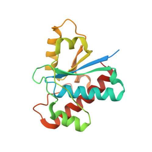
5KQL

Co-crystal structure of LMW-PTP in complex with 2-oxo-1-phenyl-2-(phenylamino)ethanesulfonic acid


Experimental Data Snapshot

  • Method: X-RAY DIFFRACTION
  • Resolution: 1.45 Å
  • R-Value Free: 0.176 
  • R-Value Work: 0.164 
  • R-Value Observed: 0.165 

wwPDB Validation   3D Report Full Report


Ligand Structure Quality Assessment 


This is version 1.1 of the entry. See complete history


Literature

Inhibition of low molecular weight protein tyrosine phosphatase by an induced-fit mechanism.

He, R.Wang, J.Yu, Z.H.Zhang, R.Y.Liu, S.Wu, L.Zhang, Z.Y.

(2016) J Med Chem 

  • DOI: https://doi.org/10.1021/acs.jmedchem.6b00993
  • Primary Citation of Related Structures:  
    5KQG, 5KQL, 5KQM, 5KQP

  • PubMed Abstract: 

    The low molecular weight protein tyrosine phosphatase (LMW-PTP) is a regulator of a number of signaling pathways and has been implicated as a potential target for oncology and diabetes/obesity. There is significant therapeutic interest in developing potent and selective inhibitors to control LMW-PTP activity. We report the discovery of a novel class of LMW-PTP inhibitors derived from sulfophenyl acetic amide (SPAA), some of which exhibit greater than 50-fold preference for LMW-PTP over a large panel of PTPs. X-ray crystallography reveals that binding of SPAA-based inhibitors induces a striking conformational change in the LMW-PTP active site, leading to the formation of a previously undisclosed hydrophobic pocket to accommodate the α-phenyl ring in the ligand. This induced-fit mechanism is likely a major contributor responsible for the exquisite inhibitor selectivity.


  • Organizational Affiliation

    Department of Medicinal Chemistry and Molecular Pharmacology, Department of Chemistry, Center for Cancer Research, and Institute for Drug Discovery, Purdue University , 720 Clinic Drive, West Lafayette, Indiana 47907, United States.


Macromolecules
Find similar proteins by:  (by identity cutoff)  |  3D Structure
Entity ID: 1
MoleculeChains Sequence LengthOrganismDetailsImage
Low molecular weight phosphotyrosine protein phosphatase178Homo sapiensMutation(s): 0 
Gene Names: ACP1
EC: 3.1.3.48 (PDB Primary Data), 3.1.3.2 (PDB Primary Data)
UniProt & NIH Common Fund Data Resources
Find proteins for P24666 (Homo sapiens)
Explore P24666 
Go to UniProtKB:  P24666
PHAROS:  P24666
GTEx:  ENSG00000143727 
Entity Groups  
Sequence Clusters30% Identity50% Identity70% Identity90% Identity95% Identity100% Identity
UniProt GroupP24666
Sequence Annotations
Expand
  • Reference Sequence
Small Molecules
Ligands 1 Unique
IDChains Name / Formula / InChI Key2D Diagram3D Interactions
6VY
Query on 6VY

Download Ideal Coordinates CCD File 
B [auth A](1~{S})-2-oxidanylidene-1-phenyl-2-phenylazanyl-ethanesulfonic acid
C14 H13 N O4 S
PCEDFPTYRADALF-ZDUSSCGKSA-N
Experimental Data & Validation

Experimental Data

  • Method: X-RAY DIFFRACTION
  • Resolution: 1.45 Å
  • R-Value Free: 0.176 
  • R-Value Work: 0.164 
  • R-Value Observed: 0.165 
  • Space Group: P 21 21 21
Unit Cell:
Length ( Å )Angle ( ˚ )
a = 34.19α = 90
b = 54.573β = 90
c = 97.515γ = 90
Software Package:
Software NamePurpose
PHENIXrefinement
HKL-3000data reduction
HKL-3000data scaling
MOLREPphasing

Structure Validation

View Full Validation Report


Ligand Structure Quality Assessment 


Entry History 

Deposition Data

Revision History  (Full details and data files)

  • Version 1.0: 2016-10-12
    Type: Initial release
  • Version 1.1: 2023-10-04
    Changes: Data collection, Database references, Derived calculations, Refinement description