5K97

Flap endonuclease 1 (FEN1) D233N with cleaved product fragment and Sm3+


Experimental Data Snapshot

  • Method: X-RAY DIFFRACTION
  • Resolution: 2.10 Å
  • R-Value Free: 0.219 
  • R-Value Work: 0.182 
  • R-Value Observed: 0.184 

wwPDB Validation   3D Report Full Report


This is version 1.4 of the entry. See complete history


Literature

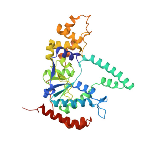
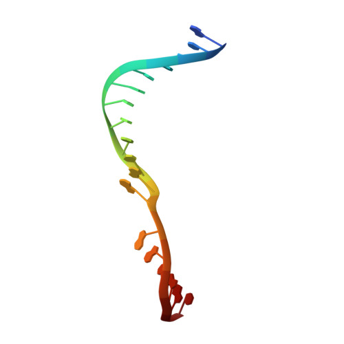
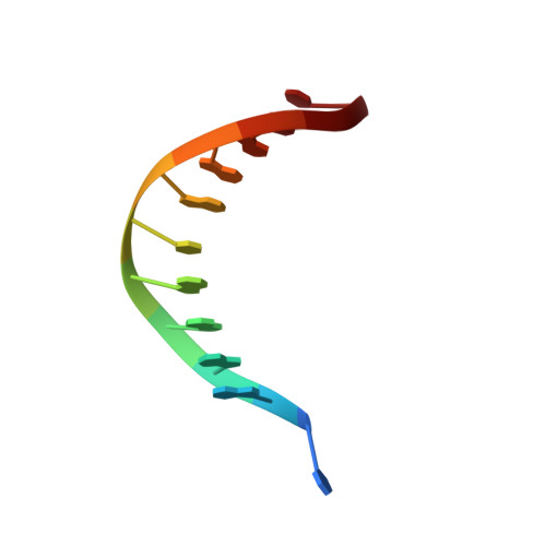
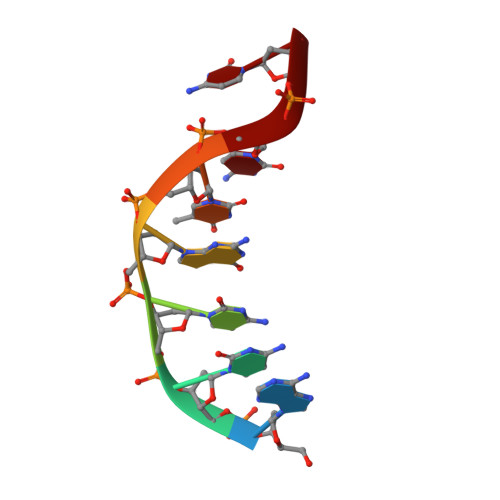
Phosphate steering by Flap Endonuclease 1 promotes 5'-flap specificity and incision to prevent genome instability.

Tsutakawa, S.E.Thompson, M.J.Arvai, A.S.Neil, A.J.Shaw, S.J.Algasaier, S.I.Kim, J.C.Finger, L.D.Jardine, E.Gotham, V.J.B.Sarker, A.H.Her, M.Z.Rashid, F.Hamdan, S.M.Mirkin, S.M.Grasby, J.A.Tainer, J.A.

(2017) Nat Commun 8: 15855-15855

  • DOI: https://doi.org/10.1038/ncomms15855
  • Primary Citation of Related Structures:  
    5K97, 5KSE, 5UM9

  • PubMed Abstract: 

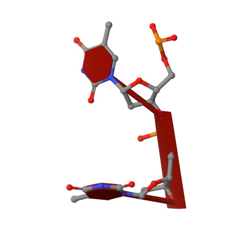
    DNA replication and repair enzyme Flap Endonuclease 1 (FEN1) is vital for genome integrity, and FEN1 mutations arise in multiple cancers. FEN1 precisely cleaves single-stranded (ss) 5'-flaps one nucleotide into duplex (ds) DNA. Yet, how FEN1 selects for but does not incise the ss 5'-flap was enigmatic. Here we combine crystallographic, biochemical and genetic analyses to show that two dsDNA binding sites set the 5'polarity and to reveal unexpected control of the DNA phosphodiester backbone by electrostatic interactions. Via 'phosphate steering', basic residues energetically steer an inverted ss 5'-flap through a gateway over FEN1's active site and shift dsDNA for catalysis. Mutations of these residues cause an 18,000-fold reduction in catalytic rate in vitro and large-scale trinucleotide (GAA) n repeat expansions in vivo, implying failed phosphate-steering promotes an unanticipated lagging-strand template-switch mechanism during replication. Thus, phosphate steering is an unappreciated FEN1 function that enforces 5'-flap specificity and catalysis, preventing genomic instability.


  • Organizational Affiliation

    Molecular Biophysics and Integrated Bioimaging, Lawrence Berkeley National Laboratory, Berkeley, California 94720, USA.


Macromolecules

Find similar proteins by:  (by identity cutoff)  |  3D Structure
Entity ID: 1
MoleculeChains Sequence LengthOrganismDetailsImage
Flap endonuclease 1341Homo sapiensMutation(s): 1 
Gene Names: FEN1RAD2
EC: 3.1
UniProt & NIH Common Fund Data Resources
Find proteins for P39748 (Homo sapiens)
Explore P39748 
Go to UniProtKB:  P39748
PHAROS:  P39748
GTEx:  ENSG00000168496 
Entity Groups  
Sequence Clusters30% Identity50% Identity70% Identity90% Identity95% Identity100% Identity
UniProt GroupP39748
Sequence Annotations
Expand
  • Reference Sequence

Find similar nucleic acids by:  Sequence   |   3D Structure  

Entity ID: 2
MoleculeChains LengthOrganismImage
DNA (5'-D(*AP*CP*TP*CP*TP*GP*CP*CP*TP*CP*AP*AP*GP*AP*CP*GP*GP*T)-3')B [auth D]18synthetic construct
Sequence Annotations
Expand
  • Reference Sequence

Find similar nucleic acids by:  Sequence   |   3D Structure  

Entity ID: 3
MoleculeChains LengthOrganismImage
DNA (5'-D(P*TP*GP*AP*GP*GP*CP*AP*GP*AP*GP*T)-3')C [auth E]11synthetic construct
Sequence Annotations
Expand
  • Reference Sequence

Find similar nucleic acids by:  Sequence   |   3D Structure  

Entity ID: 4
MoleculeChains LengthOrganismImage
DNA (5'-D(*AP*CP*CP*GP*TP*CP*C)-3')D [auth F]7synthetic construct
Sequence Annotations
Expand
  • Reference Sequence

Find similar nucleic acids by:  Sequence   |   3D Structure  

Entity ID: 5
MoleculeChains LengthOrganismImage
DNA (5'-D(P*TP*T)-3')E [auth G]5synthetic construct
Sequence Annotations
Expand
  • Reference Sequence
Small Molecules
Ligands 3 Unique
IDChains Name / Formula / InChI Key2D Diagram3D Interactions
SM
Query on SM

Download Ideal Coordinates CCD File 
F [auth A]
G [auth A]
H [auth A]
I [auth A]
K [auth A]
F [auth A],
G [auth A],
H [auth A],
I [auth A],
K [auth A],
L [auth A],
M [auth A],
N [auth A]
SAMARIUM (III) ION
Sm
DOSGOCSVHPUUIA-UHFFFAOYSA-N
EDO
Query on EDO

Download Ideal Coordinates CCD File 
O [auth A]1,2-ETHANEDIOL
C2 H6 O2
LYCAIKOWRPUZTN-UHFFFAOYSA-N
K
Query on K

Download Ideal Coordinates CCD File 
J [auth A]POTASSIUM ION
K
NPYPAHLBTDXSSS-UHFFFAOYSA-N
Experimental Data & Validation

Experimental Data

  • Method: X-RAY DIFFRACTION
  • Resolution: 2.10 Å
  • R-Value Free: 0.219 
  • R-Value Work: 0.182 
  • R-Value Observed: 0.184 
  • Space Group: P 31 2 1
Unit Cell:
Length ( Å )Angle ( ˚ )
a = 105.236α = 90
b = 105.236β = 90
c = 104.548γ = 120
Software Package:
Software NamePurpose
PHENIXrefinement
HKL-2000data collection
SCALEPACKdata scaling
PHASERphasing
PDB_EXTRACTdata extraction
HKL-2000data reduction
SCALAdata scaling

Structure Validation

View Full Validation Report


Entry History & Funding Information

Deposition Data


Funding OrganizationLocationGrant Number
National Institutes of Health/National Institute of General Medical Sciences (NIH/NIGMS)United StatesR01GM105404
National Institutes of Health/National Cancer Institute (NIH/NCI)United StatesCA92584

Revision History  (Full details and data files)

  • Version 1.0: 2017-06-28
    Type: Initial release
  • Version 1.1: 2017-07-19
    Changes: Database references
  • Version 1.2: 2017-09-27
    Changes: Author supporting evidence
  • Version 1.3: 2019-12-04
    Changes: Author supporting evidence
  • Version 1.4: 2024-02-28
    Changes: Data collection, Database references, Derived calculations