5JWP

Crystal structure of human FIH D201E variant in complex with Zn, alpha-ketoglutarate, and HIF1 alpha peptide.


Experimental Data Snapshot

  • Method: X-RAY DIFFRACTION
  • Resolution: 2.10 Å
  • R-Value Free: 0.209 
  • R-Value Work: 0.170 
  • R-Value Observed: 0.173 

wwPDB Validation   3D Report Full Report


This is version 2.2 of the entry. See complete history


Literature

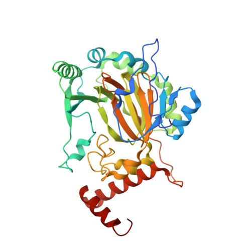
The facial triad in the alpha-ketoglutarate dependent oxygenase FIH: A role for sterics in linking substrate binding to O2 activation.

Hangasky, J.A.Taabazuing, C.Y.Martin, C.B.Eron, S.J.Knapp, M.J.

(2016) J Inorg Biochem 166: 26-33

  • DOI: https://doi.org/10.1016/j.jinorgbio.2016.10.007
  • Primary Citation of Related Structures:  
    5JWP

  • PubMed Abstract: 

    The factor inhibiting hypoxia inducible factor-1α (FIH) is a nonheme Fe(II)/αKG oxygenase using a 2-His-1-Asp facial triad. FIH activates O 2 via oxidative decarboxylation of α-ketoglutarate (αKG) to generate an enzyme-based oxidant which hydroxylates the Asn 803 residue within the C-terminal transactivation domain (CTAD) of HIF-1α. Tight coupling of these two sequential reactions requires a structural linkage between the Fe(II) and the substrate binding site to ensure that O 2 activation occurs after substrate binds. We tested the hypothesis that the facial triad carboxylate (Asp 201 ) of FIH linked substrate binding and O 2 binding sites. Asp 201 variants of FIH were constructed and thoroughly characterized in vitro using steady-state kinetics, crystallography, autohydroxylation, and coupling measurements. Our studies revealed each variant activated O 2 with a catalytic efficiency similar to that of wild-type (WT) FIH (k cat aK M(O 2 )=0.17μM -1 min -1 ), but led to defects in the coupling of O 2 activation to substrate hydroxylation. Steady-state kinetics showed similar catalytic efficiencies for hydroxylation by WT-FIH (k cat /K M(CTAD) =0.42μM -1 min -1 ) and D201G (k cat /K M(CTAD) =0.34μM -1 min -1 ); hydroxylation by D201E was greatly impaired, while hydroxylation by D201A was undetectable. Analysis of the crystal structure of the D201E variant revealed steric crowding near the diffusible ligand site supporting a role for sterics from the facial triad carboxylate in the O 2 binding order. Our data support a model in which the facial triad carboxylate Asp 201 provides both steric and polar contacts to favor O 2 access to the Fe(II) only after substrate binds, leading to coupled turnover in FIH and other αKG oxygenases.


  • Organizational Affiliation

    Department of Chemistry, University of Massachusetts, Amherst, United States.


Macromolecules
Find similar proteins by:  (by identity cutoff)  |  3D Structure
Entity ID: 1
MoleculeChains Sequence LengthOrganismDetailsImage
Hypoxia-inducible factor 1-alpha inhibitor352Homo sapiensMutation(s): 1 
Gene Names: HIF1ANFIH1
EC: 1.14.11.30 (PDB Primary Data), 1.14.11 (PDB Primary Data)
UniProt & NIH Common Fund Data Resources
Find proteins for Q9NWT6 (Homo sapiens)
Explore Q9NWT6 
Go to UniProtKB:  Q9NWT6
PHAROS:  Q9NWT6
GTEx:  ENSG00000166135 
Entity Groups  
Sequence Clusters30% Identity50% Identity70% Identity90% Identity95% Identity100% Identity
UniProt GroupQ9NWT6
Sequence Annotations
Expand
  • Reference Sequence

Find similar proteins by:  Sequence   |   3D Structure  

Entity ID: 2
MoleculeChains Sequence LengthOrganismDetailsImage
Hypoxia-inducible factor 1-alpha19Homo sapiensMutation(s): 1 
UniProt & NIH Common Fund Data Resources
Find proteins for Q16665 (Homo sapiens)
Explore Q16665 
Go to UniProtKB:  Q16665
PHAROS:  Q16665
GTEx:  ENSG00000100644 
Entity Groups  
Sequence Clusters30% Identity50% Identity70% Identity90% Identity95% Identity100% Identity
UniProt GroupQ16665
Sequence Annotations
Expand
  • Reference Sequence
Small Molecules
Ligands 4 Unique
IDChains Name / Formula / InChI Key2D Diagram3D Interactions
AKG
Query on AKG

Download Ideal Coordinates CCD File 
C [auth A]2-OXOGLUTARIC ACID
C5 H6 O5
KPGXRSRHYNQIFN-UHFFFAOYSA-N
SO4
Query on SO4

Download Ideal Coordinates CCD File 
F [auth A],
G [auth A],
H [auth A],
I [auth A]
SULFATE ION
O4 S
QAOWNCQODCNURD-UHFFFAOYSA-L
GOL
Query on GOL

Download Ideal Coordinates CCD File 
D [auth A]GLYCEROL
C3 H8 O3
PEDCQBHIVMGVHV-UHFFFAOYSA-N
ZN
Query on ZN

Download Ideal Coordinates CCD File 
E [auth A]ZINC ION
Zn
PTFCDOFLOPIGGS-UHFFFAOYSA-N
Experimental Data & Validation

Experimental Data

  • Method: X-RAY DIFFRACTION
  • Resolution: 2.10 Å
  • R-Value Free: 0.209 
  • R-Value Work: 0.170 
  • R-Value Observed: 0.173 
  • Space Group: P 41 21 2
Unit Cell:
Length ( Å )Angle ( ˚ )
a = 86.22α = 90
b = 86.22β = 90
c = 149.903γ = 90
Software Package:
Software NamePurpose
HKL-2000data collection
HKL-2000data scaling
REFMACrefinement
PDB_EXTRACTdata extraction
HKL-2000data reduction
PHASERphasing

Structure Validation

View Full Validation Report


Entry History & Funding Information

Deposition Data


Funding OrganizationLocationGrant Number
National Institutes of Health/National Institute of General Medical Sciences (NIH/NIGMS)United StatesT32-GM008515

Revision History  (Full details and data files)

  • Version 1.0: 2016-11-23
    Type: Initial release
  • Version 2.0: 2017-09-27
    Changes: Author supporting evidence, Database references, Polymer sequence
  • Version 2.1: 2019-12-25
    Changes: Author supporting evidence
  • Version 2.2: 2023-09-27
    Changes: Data collection, Database references, Refinement description