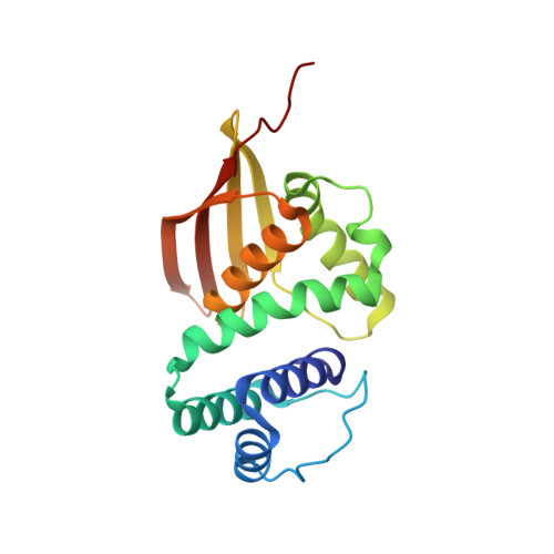
Structural and Functional Evidence Indicates Selective Oxygen Signaling in Caldanaerobacter subterraneus H-NOX.
Hespen, C.W., Bruegger, J.J., Phillips-Piro, C.M., Marletta, M.A.(2016) ACS Chem Biol 11: 2337-2346
- PubMed: 27328180
- DOI: https://doi.org/10.1021/acschembio.6b00431
- Primary Citation of Related Structures:
5JRU, 5JRV, 5JRX - PubMed Abstract:
Acute and specific sensing of diatomic gas molecules is an essential facet of biological signaling. Heme nitric oxide/oxygen binding (H-NOX) proteins are a family of gas sensors found in diverse classes of bacteria and eukaryotes. The most commonly characterized bacterial H-NOX domains are from facultative anaerobes and are activated through a conformational change caused by formation of a 5-coordinate Fe(II)-NO complex. Members of this H-NOX subfamily do not bind O2 and therefore can selectively ligate NO even under aerobic conditions. In contrast, H-NOX domains encoded by obligate anaerobes do form stable 6-coordinate Fe(II)-O2 complexes by utilizing a conserved H-bonding network in the ligand-binding pocket. The biological function of O2-binding H-NOX domains has not been characterized. In this work, the crystal structures of an O2-binding H-NOX domain from the thermophilic obligate anaerobe Caldanaerobacter subterraneus (Cs H-NOX) in the Fe(II)-NO, Fe(II)-CO, and Fe(II)-unliganded states are reported. The Fe(II)-unliganded structure displays a conformational shift distinct from the NO-, CO-, and previously reported O2-coordinated structures. In orthogonal signaling assays using Cs H-NOX and the H-NOX signaling effector histidine kinase from Vibrio cholerae (Vc HnoK), Cs H-NOX regulates Vc HnoK in an O2-dependent manner and requires the H-bonding network to distinguish O2 from other ligands. The crystal structures of Fe(II) unliganded and NO- and CO-bound Cs H-NOX combined with functional assays herein provide the first evidence that H-NOX proteins from obligate anaerobes can serve as O2 sensors.
Organizational Affiliation:
Department of Molecular and Cell Biology, University of California-Berkeley , 356 Stanley Hall, Berkeley, California 94720-3220, United States.